
In the above article (Shafiei K, Luther E, Archie M, Gulick J, Fowler MR. J Am Board Fam Pract 16:555–9), the four figures and figure legends were mismatched. They are printed correctly below. We regret this error and apologize for any confusion or inconvenience it may have caused.

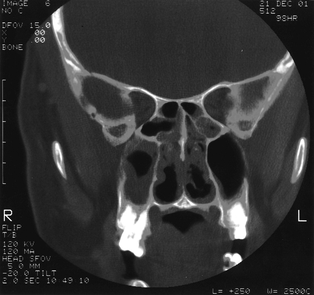
Figure 1.
Coronal CT of sinuses demonstrating bilateral ethmoid and maxillary sinus disease.

Figure 2.
Chest CT demonstrating soft tissue mass in left upper lobe abutting the pleura.
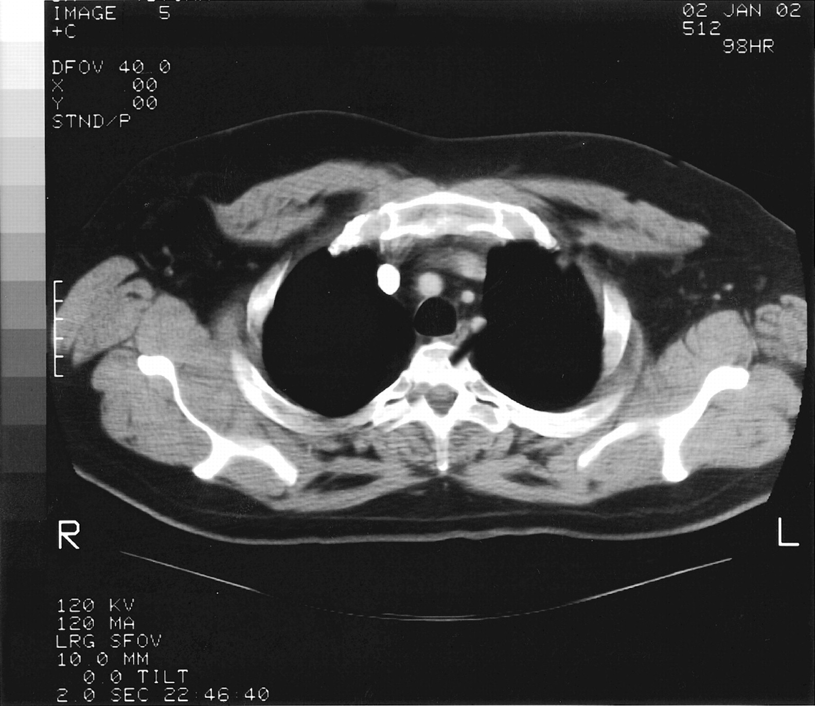
Figure 3.
Chest CT demonstrating small noncalcified lesions in the left upper lobe.

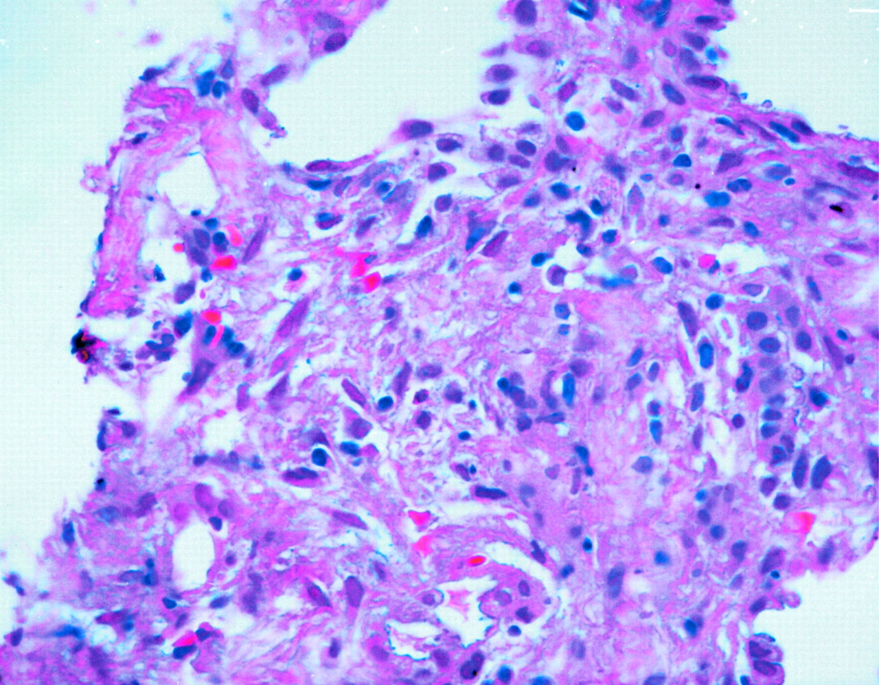
Figure 4.
Focus of granulomatous inflammation, lung (hematoxylin and eosin; original magnification, 400×).


Jump to section
Related Articles
Cited By...
- No citing articles found.