
Article Figures & Data
Figures
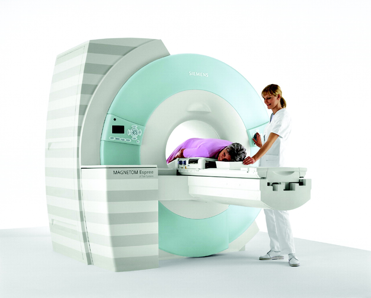
- Figure 1.
MR scanning of a patient using a dedicated breast coil
Tables
Applications MEDLINE Search Terms Staging of breast cancer (including determining involvement of pectoral musculature) Magnetic resonance imaging AND breast cancer staging Determination of recurrent/residual disease after treatment Magnetic resonance imaging AND breast cancer AND residual disease Determination of occult breast cancer (especially in patients with negative mammograms) Magnetic resonance imaging AND breast cancer AND unknown primary Possible use as a screening tool in patients at high risk for breast cancer Magnetic resonance imaging AND breast cancer AND high risk Study Type of Study Study Population N Major Findings/Comments Boetes et al.23 Retrospective Patients with invasive lobular carcinoma treated surgically 34 (36 cases of breast cancer) False negative rate for MRI was 0% compared with 3% and 14% for US and XRM, respectively; when 2 radiologists retrospectively reviewed the exam results, the percentage of correctly identified size of cancer was 47% and 75% for MRI (r = 0.81, P < .01); this was the most accurate and most highly correlated method Schelfout et al.17 Prospective Women with lesions on XRM, US, and/or CBE 212 96%, 37%, and 41% of multifocal disease was detected by MRI, XRM, and US, respectively; 95%, 18%, and 9% of multicentric disease was detected by MRI, XRM, and US, respectively; 100% of bilateral breast cancers were seen on MRI; 56% of bilateral breast cancers were seen on XRM and US Van Goethem et al.24 Prospective Patients with dense breasts planning to undergo surgery 67 65/67 patients had breast cancer confirmed pathologically; MRI was 98% sensitive for initial lesion compared with 83% and 70.8% for XRM and US, respectively; extent of cancer was underestimated by 12.5% of MRI results as opposed to 37% and 40% for XRM and US results, respectively; multifocal/multicentric disease was picked up 100% of the time by MRI as opposed to 35% and 30% of XRM and US, respectively Bedrosian et al.18 Retrospective Patients diagnosed with invasive breast cancer who had MRI preoperatively 267 MRI was 95% sensitive for detecting primary breast cancer; planned management of 26% of patients (N = 69) were changed due to MRI results—in 49/69 (71%) of the patients, postsurgical pathology confirmed that the change in management was appropriate Rieber et al.28 Prospective Patients suspected of having breast malignancy based on results of XRM, US, or CBE 43 Sensitivity of MR mammography for diagnosis of primary cancer, contralateral cancer, and multifocal disease was 100%, 100%, and 95.2%; similar values were 93%, 100%, and 92.5% for PET, respectively Fischer et al.20 Prospective Patients with breast abnormalities after XRM, CBE, PE, and US 463 66 patients (14.3%) had their planned therapy accurately changed as a result of MRI of the breast with 16 patients (3.5%) undergoing unneeded open biopsy Esserman et al.21 Prospective Patients with breast cancer with planned surgical correction 57 (58 cases of breast cancer) Preoperative MRI identified degree of disease accurately in 54/58 cases of breast cancer; anatomic detail identified on MRI was accurate 98% of the time whereas anatomic detail identified on XRM was accurate in 55% of cases Rodenko et al.22 Retrospective Patients with infiltrating lobular carcinoma who had MRI and XRM performed preoperatively 20 Postoperative pathology correlated with preoperative MRI findings in 85% of patients; The correlation with preoperative XRM was 32% (P < .0001) Study Type of Study Study Population N Major Findings/Comments Yeh et al.25 Prospective Patients with stage IIb/III breast cancer undergoing neoadjuvant chemotherapy (doxorubicin and paclitaxel) 31 Correlation with pathology Equal to pathology/underestimate/overestimate MRI: 71%/23%/6% XRM: 26%/52%/23% US: 35%/52%/13% MRI was best correlated with pathology (P < .002) Chen et al.26 Prospective Patients with locally advanced breast cancer receiving neoadjuvant therapy (adriamycin, cytoxan, and paclitaxel) 15 MRI tended to overestimate tumor response; for partial non-responders MRI correlated with pathology 0% of the time compared with 17% for CBE and 83% for PET; however, for responders, MRI correlated with pathology 90% of the time, with 70% correlation for CBE and 90% for PET; MRI size measurements of residual tumors did correlate with pathology (coefficient 0.7 compared with −0.06 for CBE) Denis et al.27 Prospective Patients with locally advanced breast cancer receiving neoadjuvant therapy with either 5–5-fluorouracil/epirubicin/ cyclophosphamide; docetaxel only, or docetaxel with epirubicin 40 Correlation coefficient of MRI measurements with pathology: 5-fluorouracil/E/C: 0.89, DXL: 0.64, DXL/E: 0.16 MRI overestimated tumor response in DXL-based groups, which was believed to be due to antiangiogenic effect of DXL, which would impair MRI contrast enhancement; this was supported by the fact that DXL-based groups had residual disease of microscopic nests of tumor cells on pathology as opposed to single nodular lesions Tozaki et al.28 Prospective Patients with locally advanced breast cancer (IIb/III) undergoing neoadjuvant chemotherapy 19 Accuracy (deviation from pathology of less than 2 mm) of late phase CT and MRI scans (scan 4 minutes after contrast injection) compared with pathology was up to 90% for DCIS and replaced (diffuse pre-NAC contrast enhancement) lesions and 88% for nonreplaced (localized CE) lesions; early phase scans were 0% accurate for DCIS/replaced and 75% accurate for nonreplaced lesions Bodini et al.29 Prospective Patients with T2 to 4, N0 M0 breast cancer treated with 3 to 4 cycles of epirubicin 79 Clinical response correlation with pathology correlation coefficients were 0.72 for MRI and 0.68 for CBE Warren et al.30 Retrospective Patients undergoing neoadjuvant therapy 67 MRI was more sensitive and specific for assessment of complete or partial response (100% and 80%) than conventional assessment methods (CAM), including XRM, US, and CBE (98% and 50%); agreement with pathology was marginally higher in MRI compared with CAM (81% compared to 68%, P = .09); MRI increased diagnostic knowledge in 70% of patients, and increased diagnostic confidence in 52%; however, MRI did not change treatment plan, decreased confidence in 20% of patients, and decreased knowledge in 17%; MRI tended to overestimate response Lee et al.31 Retrospective Patients who had excisional biopsy who required definitive surgery, eg, because of positive margins/residual disease and were candidates for breast conservation 80 MRI sensitivity and specificity for residual disease was 61.2% and 69.7%, respectively; additional lesions were detected by imaging of which 10 were only seen on MRI; of these, 5 were benign, 5 were malignant; MRI changed management of patients in 29% of the cases, because of additional lesions found; whether this led to improved patient outcomes is not known Rosen et al.32 Retrospective Patients with locally advanced breast cancer treated with neoadjuvant therapy (paclitaxel, doxorubicin, and breast hyperthermia) 21 Correlation with pathology equal to pathology/underestimate/overestimate MRI: 57%/10%/33% correlation coefficients were 0.74 for MRI and 0.65 (statistically nonsignificant trend); in contrast to some of the other studies, MRI tended to overestimate the residual tumor (ie, underestimate response) Hwang et al.33 Retrospective Patients with histologically confirmed diagnosis of DCIS 51 MRI was 97% sensitive and 58% specific for detecting residual disease compared with histology; the sensitivity was significantly better than the sensitivity of XRM for residual disease; in addition MRI had a significantly higher NPV compared to XRM Belli et al.34 Prospective Patients with locally advanced breast cancer being treated with neoadjuvant chemotherapy 45 MRI had a 90.2% sensitivity and a 100% specificity for residual disease Cheung et al.35 Prospective Patients with locally advanced breast cancer being treated with neoadjuvant chemotherapy 33 Residual tumor on MRI correlated with microscopic findings (correlation coefficient = 0.982, P < .001) Partridge et al.36 Prospective Patients with invasive breast cancer undergoing neoadjuvant therapy with doxorubicin and cyclophosphamide 52 Decreased tumor enhancement prechemotherapy and postchemotherapy (210% vs 166%, P < .001); MRI had correlation coefficient of 0.89 compared with pathology whereas clinical examination had coefficient of 0.60 Kawashima et al.37 Prospective Patients who underwent excisional biopsy of breast lesion 50 MRI in detection of residual disease: sensitivity 66%; specificity 81%; PPV 72%; NPV 83%; accuracy 63% Drew et al.38 Prospective Patients with locally advanced breast cancer receiving neoadjuvant therapy 17 Dynamic CE MRI was 100% accurate in assessing residual disease; CBE and XRM were not sensitivity/specificity/PPV/NPV CBE: 50%/86%/83%/55% XRM: 90%/57%/75%/80% Weatherall et al.39 Retrospective Patients with breast cancer with chemotherapy prior to surgery 20 MRI demonstrated a correlation with pathology (coefficient = 0.93); coefficients for CBE and XRM were 0.72 and 0.63, respectively Frei et al.40 Retrospective Patients with excisional biopsy 68 MRI sensitivity/specificity/PPV/NPV >7 days postbiopsy: 89%/52%/81%/69% >14 days postbiopsy: 88%/58%/82%/69% >21 days postbiopsy: 91%/69%/88%/75% >28 days postbiopsy: 92%/75%/92%/75% >35 days postbiopsy: 95%/75%/91%/86% >42 days postbiopsy: 94%/75%/89%/86%The peak values for PPV and plateau for specificity occurs at >28 days, after which the improvement is not as much; therefore, this may be the best time to perform the MRI as the PPV of positive margins in this study was 69% compared to 92% for MRI at day 28, which may lead to breast-conserving surgery Trecate et al.41 Prospective Patients with locally advanced breast cancer receiving chemotherapy 30 MRI: sensitivity: 96%; specificity: 75%; PPV: 92.5%; NPV 66%; accuracy 90% Orel et al.42 Prospective Patients who underwent excisional biopsy 47 MRI had PPV of 82% and NPV of 61%; false negatives possibly secondary to postsurgical changes Soderstrom et al.43) Prospective Patients who underwent excisional biopsies 19 MRI had an 84% accuracy in determining whether residual tumor was present in patients postexcisional biopsy Study Type of Study Study Population N Mean Age/Range Major Findings/Comments Olson et al.45 Prospective Women with unknown primary and metastatic axillary adenocarcinoma; initial XRM, US, or PE was not diagnostic of malignancy 40 58 (+) MRI correlated with breast tumor in 81% of cases overall; initial XRM, US, or PE was not diagnostic of malignancy Henry-Tillman RS et al. (Dec 1999) (54) Retrospective Patients with unknown primary, (+) axillary/supraclavicular lymphadenopathy, (−) XRM/CBE, and (+) MRI 10 36 to 68 MRI was 100% accurate when correlated with pathology; 80% (N = 8) were (+) and primary breast CA was confirmed; 20% (N = 2) were negative and other primaries (ovarian CA and lymphoma) were identified Orel SG et al. (Aug 1999) (55) Prospective Patients with (+) axillary node metastasis, negative PE and XRM, and unknown primary 22 49 Breast cancer was detected as the primary in 86% (N = 19) of cases; 17 were confirmed by pathology; 2 resolved on MRI follow-up during chemotherapy Study Type of Study Study Population N Mean Age/Range Major Findings/Comments Leach et al.55 Prospective Women at high risk for breast cancer (BRCA 1, 2, or TP53 mutation, 1st degree relative with mutation, family history of breast/ovarian CA, family history of Li-Fraumeni syndrome) 649 31 to 55 Sensitivity for CE MRI and XRM was 77% and 40%, respectively; specificity for CE MRI and XRM was 81% and 93%, respectively; sensitivity and specificity for both methods combined was 94% and 77%, respectively; sensitivity of CE MRI for BRCA 1 and BRCA 2 were 92% and 58%, respectively Lehman et al.56 Prospective Women ≥25 with high familial or genetic risk of breast cancer 367 40 1.1% of patients screened with MRI had breast cancer by pathology as opposed to 0.3% by XRM (difference not statistically significant); PPV of biopsies performed as a result of MRI was 17% (4/24); PPV of biopsies performed as a result of XRM was 25% (1/4) Sim et al.58 Retrospective Women who were at least at 15% greater risk for breast cancer (Claus model) 179 Sensitivity for MRI, XRM, and US were 93.3%, 83.3%, and 53.9%, respectively; specificity for MRI, XRM, and US were 63.6%, 65.5%, and 85.7%, respectively; the sensitivity and specificity for combined XRM and US was 92.9% and 62.5%, respectively Kriege et al.50 Prospective Women who were at least at 15% greater risk for breast cancer due to familial or genetic factors (Claus model) 1909 40 Sensitivity for MRI in detecting invasive breast cancer as opposed to CBE and XRM was 79.5%, 17.9%, and 33.3%, respectively; specificity for MRI, CBE, and XRM was 89.8%, 98.1%, and 95.0%, respectively Morris et al.54 Retrospective Women at high risk for breast cancer (past history, family history, LCIS, atypia) with negative XRM 367 50 4% of screened patients had breast cancer by pathology (57% were DCIS); percentage was higher in those women with past and family history (8%), PPV 50%; PPV of biopsy was 24% Warner et al.49 Prospective Women who were carriers of BRCA 1 and/or BRCA 2 236 25 to 65 Sensitivity for MRI as opposed to XRM, US, and CBE were 77%, 36% (P = .02), 33% (P = .006), and 9.1%, respectively; specificity for MRI, XRM, US, and CBE were 95.4%, 99.8%, 96%, and 99.3%, respectively; when all modalities were combined there was a sensitivity of 95% as opposed to 45% with XRM and CBE combined Podo et al.57 Prospective Women with confirmed BRCA 1 or BRCA 2 mutations or with a 1 in 2 chance of having the mutation 105 55.3 8 breast cancers were identified (all by CE MRI); only 1 was detected by XRM and US; total incidence of breast cancer was 7.6%


