
Abstract
Coccidioidomycosis is a fungal infection endemic to the Southwestern United States. Extrapulmonary Coccidioides immitis infections are uncommon and occur more frequently in immunocompromised individuals. There is often a delay in diagnosis and treatment due to the chronic, indolent nature of these infections. The clinical presentation is often nonspecific, and includes joint pain, erythema, or localized swelling. Therefore, these infections may only be identified after initial treatment failure and further workup is pursued. The majority of reported cases of coccidioidomycosis involving the knee have had intra-articular involvement or extension. This report describes a rare case of peri-articular Coccidioides immitis abscess of the knee that does not communicate with the joint in a healthy patient. This case illustrates the low threshold needed for additional testing, such as fluid or tissue sampling of joint-related fluid collections if the etiology is unclear. A high index of suspicion is prudent to avoid diagnostic delay, particularly for individuals who either reside in or travel to endemic areas.
Case
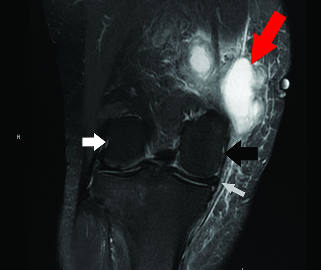
A previously healthy, 64-year-old female presented with 6 weeks of atraumatic, right medial knee pain and localized fluid collection. She attributed the new swelling to a recent increase in her incline treadmill exercise. Her medical history was only significant for a right upper lobe lung nodule, which had been an incidental finding and stable through serial imaging approximately 2 years prior. She otherwise had no immunocompromising medical conditions. Aside from the reported right medial knee pain and nontender, focal medial knee swelling, the remainder of her knee examination was normal, and she had no other symptoms or complaints. After 2 visits in Primary Care clinic (1 of which was virtual), she was seen in Primary Care Sports Medicine clinic and underwent aspiration of the fluid collection, which showed a turbid, brown fluid. The aspirate was sent for bacterial and fungal cultures (routinely performed together at our institution’s laboratory), cell count with differential, Gram stain and crystal analysis. The culture of the fluid identified fungal elements 2 days after aspiration, and eventually grew Coccidioides immitis 9 days after collection. Subsequently, she was referred to Orthopedic and Infectious Disease specialists. Magnetic resonance imaging (MRI) of the knee demonstrated a 6-cm × 4.3-cm × 5-cm abscess adjacent to the medial collateral ligament (Figure 1). This abscess did not communicate with the joint. Initially, the orthopedic specialist performed a therapeutic aspiration, and the infectious disease specialist prescribed 400 milligrams of oral fluconazole twice daily for 2 weeks. Despite these interventions, the fluid reaccumulated within 2 weeks, and the patient required an open incision and drainage in the operating room. Aside from the previously identified stable right pulmonary nodule, further imaging of the brain and thorax did not reveal additional lesions concerning for disseminated infection. After surgical treatment, she was continued on oral fluconazole 400 milligrams daily, with anticipation to continue this treatment for 2 years total. The fluid has not recurred since that time, and she has been able to return to her usual exercise without difficulty.

T2-wighted magnetic resonance image of the patient’s right knee. Notes: The medial and lateral femoral condyles are labeled as the black and white arrows, respectively. The 6-cm × 4.3-cm × 5-cm extra-articular abscess (red arrow) is adjacent to the medial collateral ligament (gray arrow).
Discussion
Coccidioidomycosis is a fungal infection endemic to the Southwestern United States. It is caused by inhalation of the spores of Coccidioides immitis. While 60% of infections are asymptomatic, the remaining 40% typically result in mild, self-limited complaints, often due to pulmonary disease.1,2 In most cases, there is known travel within an endemic area. Disseminated extrapulmonary infections are rare, occurring in less than 1% of new cases annually.1,3,4 Patients at highest risk for disseminated infections include those of African American or Filipino descent,5 or who have immunosuppressing medical conditions, such as diabetes, tobacco use, autoimmune conditions, cardiopulmonary disease and advanced age.4
Among disseminated extrapulmonary Coccidioides infections, the spine and knee are the most commonly affected musculoskeletal structures.6–7 These infections occur through hematogenous spread or by direct extension through a bone or surrounding soft tissues. This results in a variety of clinical conditions depending on the structure involved, including synovitis, arthritis, osteomyelitis, tenosynovitis, tendinitis, abscesses, or cutaneous infections. The patient typically presents with joint pain, effusion, erythema, or localized swelling.8 Although the majority of cases involving the knee affect intra-articular structures, there are rare cases of infections localized to the extra-articular space, including popliteal cysts9–10 and superficial abscesses.8 Such infections, similar to the present case, may result in localized swelling with few other findings.
It may take years for extrapulmonary infections to be correctly identified due to their indolent nature. The clinical course is often chronic and fluctuating, and may mimic other degenerative conditions, such as osteoarthritis.11 Often, these fungal infections are identified only after an initial course of treatment has failed and further investigation is performed. A preceding pulmonary illness may provide a historic clue to the initial Coccidioides infection, however signs of disseminated disease may take years to manifest.12–13 Therefore, a high index of suspicion may be needed to correctly diagnose these infections.
Coccidioides infections can be diagnosed based on serologic testing, histopathologic examination or by culture.6,14,15 Nonspecific laboratory findings include eosinophilia2 and elevated inflammatory markers (such as the erythrocyte sedimentation rate).7 Serologic (antibody) tests are most commonly used as initial screening, as this is noninvasive and often returns more rapidly than cultures.16–17 IgM antibodies may become positive in the first 1 to 3 weeks of the infection, thereby allowing for detection of acute disease.2,16 IgG antibody levels typically develop later and can be used to indicate the degree of infection.2,16,18 The sensitivity and specificity of these tests vary according to the laboratory and the specific testing method that is used.2,16 Immunocompromised patients may not yield the same antibody response as an immunocompetent patient, however, contributing to diagnostic delay.2,19
Definitive diagnosis often requires histopathologic examination or culture of the affected tissue, as these are considered the gold standard for diagnosis.17 Microscopy of extrapulmonary lesions can demonstrate characteristic spherules on fungal stain.2,17 A specific request to evaluate for fungal organisms may be required if the evaluating laboratory does not routinely test for fungal organisms when performing fluid cultures. Not all synovial fluid cultures successfully grow Coccidioides, however.6 In such cases, arthroscopic examination and synovial tissue biopsy for histopathology may be required. If there is a high index of suspicion and initial cultures are nondiagnostic, exoantigen and DNA probe testing can also be used to diagnose Coccidioides infections.2,12,17 Serial testing (whether by serology, cytology, histopathology or culture) every several weeks can increase sensitivity if the initial tests are negative and clinical suspicion remains high.17
Diagnostic imaging shows nonspecific findings. Radiographs are typically used as the initial imaging modality and may show effusion, periarticular bony destruction, lytic bone lesions or degenerative changes.20 CT and MRI provide further information about soft tissue and may show joint effusion, hypertrophied synovium, abscesses, bony edema or destructive bony lesions.20 Radionuclide scanning and bone scintigraphy can be used to detect disseminated disease; however MRI is considered more sensitive for detecting early disease within a joint.7
The majority of these infections require a combination of both medical and surgical treatments, which should be made in consultation with infectious disease and orthopedic specialists. In some cases, such as infection limited to the synovium, the patient may require medical treatment only.11 Antifungal therapy is recommended for all cases of extrapulmonary soft tissue coccidioidomycosis. Triazole fungicides such as fluconazole or itraconazole, are the preferred antifungal medications for Coccidioides infections. Amphotericin B is an alternative therapy that can be used in the case of azole failure or in cases of severe, rapidly progressing infections which necessitate more timely treatment.18 Otherwise, amphotericin B is often avoided due to its toxicity concerns.
The decision to pursue surgical intervention is based on the location of the infection, the structures affected, its potential to compromise neighboring structures (such as regional nerves), or cases refractory to medical management.21 Surgical treatment typically includes joint debridement, synovectomy or resection of diseased tissues.6,8,11,12,22⇓–24 The ultimate goal of surgery is to reduce necrotic or diseased tissue to healthy margins.8 Severe or rapidly progressing neurologic symptoms may indicate the need for urgent surgical intervention.18 Regardless of surgical outcomes, antifungal medications are nearly always used before and after surgical intervention.21
Long-term recovery and duration of antifungal treatment varies from months to lifelong suppression depending on each individual case and level of immunocompetency.6,11,22⇓–24 Recovery is variable among cases, as some patients return to activity without difficulty, as in our case, while others have persistent symptoms and require additional interventions.
Conclusion
This case illustrates that Coccidioides immitis infections can cause peri-articular infections that do not communicate with the joint in healthy patients. Coccidioides infections can be a diagnostic challenge due to their indolent nature.11 Additional testing, such as fluid or tissue sampling, may be needed to secure the diagnosis in patients who have risk factors for this infection. Serologic and histopathologic testing should be considered in patients who are not improving with conservative management. Many patients require a combination of medical and surgical interventions once diagnosed. Therefore, a multidisciplinary team, including infectious disease and orthopedic specialists, should be utilized to help guide treatment.
Notes
This article was externally peer reviewed.
Funding: None.
Conflicts of interest: None.
To see this article online, please go to: http://jabfm.org/content/36/2/376.full.
- Received for publication July 6, 2022.
- Revision received October 24, 2022.
- Revision received October 31, 2022.
- Accepted for publication November 2, 2022.


