
Main menu

  • HOME
  • ARTICLES
    • Current Issue
    • Ahead of Print
    • Archives
    • Abstracts In Press
    • Special Issue Archive
    • Subject Collections
  • INFO FOR
    • Authors
    • Reviewers
    • Call For Papers
    • Subscribers
    • Advertisers
  • SUBMIT
    • Manuscript
    • Peer Review
  • ABOUT
    • The JABFM
    • The Editing Fellowship
    • Editorial Board
    • Indexing
    • Editors' Blog
  • CLASSIFIEDS
  • Other Publications
    • abfm

User menu

Search

  • Advanced search
American Board of Family Medicine
  • Other Publications
    • abfm
American Board of Family Medicine

American Board of Family Medicine

Advanced Search

  • HOME
  • ARTICLES
    • Current Issue
    • Ahead of Print
    • Archives
    • Abstracts In Press
    • Special Issue Archive
    • Subject Collections
  • INFO FOR
    • Authors
    • Reviewers
    • Call For Papers
    • Subscribers
    • Advertisers
  • SUBMIT
    • Manuscript
    • Peer Review
  • ABOUT
    • The JABFM
    • The Editing Fellowship
    • Editorial Board
    • Indexing
    • Editors' Blog
  • CLASSIFIEDS
  • JABFM on Bluesky
  • JABFM On Facebook
  • JABFM On Twitter
  • JABFM On YouTube
Research ArticleBrief Report

Therapeutic Dermoscopy to Facilitate Detection and Extraction of Foreign Bodies

Sody A. Naimer
The Journal of the American Board of Family Medicine May 2017, 30 (3) 374-376; DOI: https://doi.org/10.3122/jabfm.2017.03.160365
Sody A. Naimer
From the Department of Family Medicine, Siaal Research Center for Family Medicine and Primary Care, Division of Community Health, Faculty of Health Sciences, Ben-Gurion University of the Negev, Beer-Sheva, Israel; and the Eilon Moreh Clinic, Clalit Health Services, Shomron District, Lev Shomron, Israel.
MD
  • Find this author on Google Scholar
  • Find this author on PubMed
  • Search for this author on this site
  • Article
  • Figures & Data
  • References
  • Info & Metrics
  • PDF
Loading

Abstract

Foreign bodies are occasionally seen by family physicians. Plantar foreign bodies in particular pose a special challenge because they involve weight-bearing regions that are difficult to access. If left undetected long enough, these may lead to hospitalization, surgery, or even longstanding complications such as tumors, contractures, infections, and chronic ulcers. Dermoscopy of the cutaneous surface allows early detection of indwelling foreign bodies with a far greater degree of accuracy than with the naked eye. Furthermore, use of a polarized dermatoscope provides ideal illumination and 3-dimensional visualization of the involved site, and facilitates extraction of the penetrating object. This technique could be used for similar injuries involving other body surfaces.

  • Dermoscopy
  • Foreign Bodies
  • Family Physicians

Foreign bodies are occasionally seen by family physicians. Plantar foreign bodies in particular pose a great challenge because they involve weight-bearing surfaces in an awkward location.1 Despite the seemingly benign character of an injury caused by stepping on a thorn or an unrecognized sliver of glass finding its way deep into the thick epidermal layer of the plantar skin, such events may lead to hospitalization, surgery, or even long-term complications such as tumors, contractures, infections, and chronic ulcers.2⇓⇓⇓–6 If ignored, imaging may be needed to locate the penetrating object.7,8 It is assumed that clinicians would welcome a technique enabling prompt diagnosis and removal of most foreign bodies before the onset of difficult-to-treat complications.

The dermatoscope has attained growing popularity among family physicians. Its status has been quoted by specialists as acquiring “a role similar to the stethoscope of general practitioners, becoming an irreplaceable clinical tool for dermatologists.”9 This instrument offers a marked contribution in performing challenging therapeutic procedures. It has already been reported to facilitate the identification of obscure conditions such as the detection and extraction of unidentified corneal foreign bodies or buried sutures.10,11

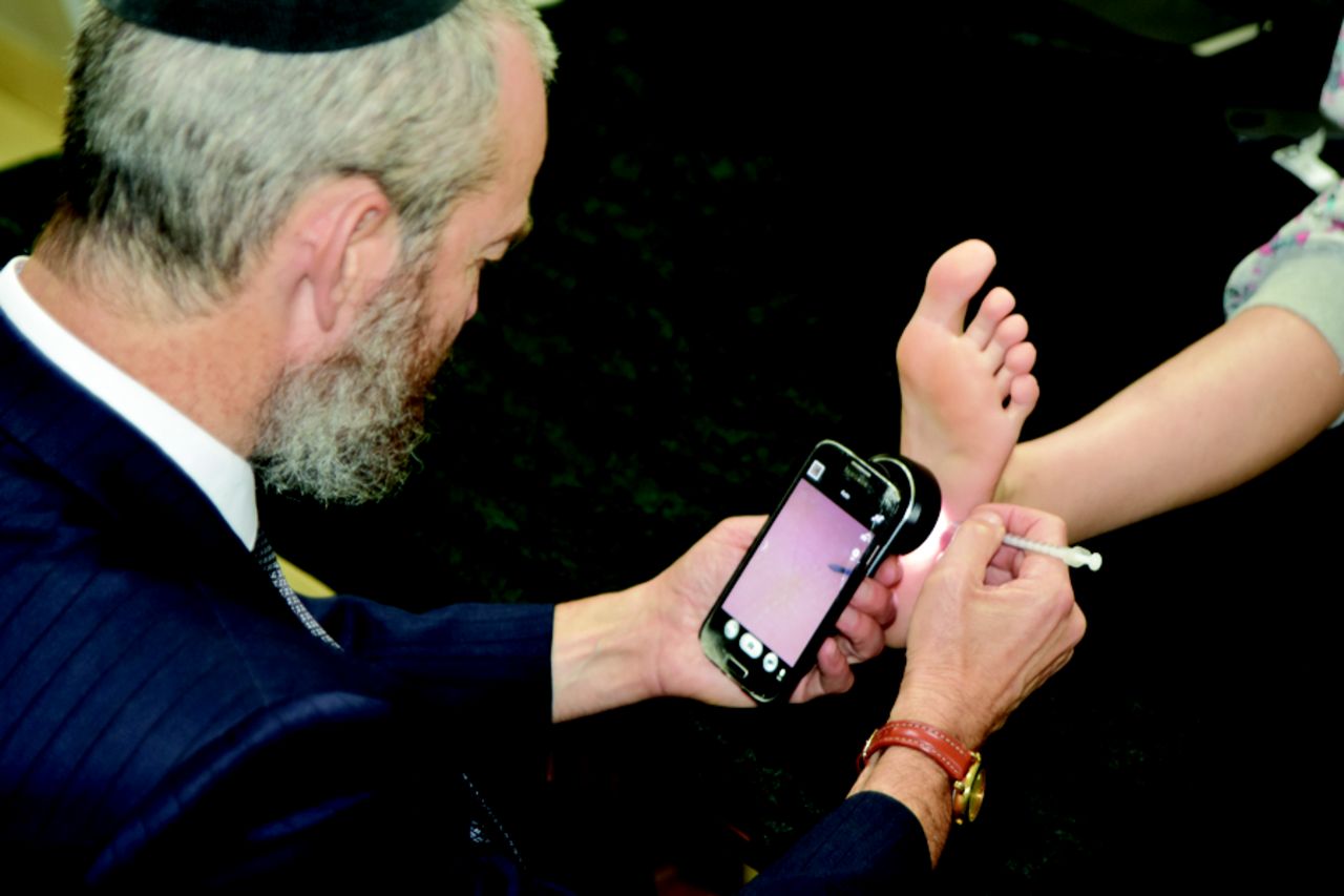
Dermoscopy is recommended whenever a lesion can be characterized better with ideal illumination coupled with magnified visualization. The benefit conferred by a polarized dermatoscope, beyond the original episcope, is excellent focal accommodation without actual adherence to the surface being examined, which opens the possibility to perform procedures during observation of the involved area. This sets the stage for “therapeutic dermoscopy.” An adapter is available that attaches a standard smartphone to the eyepiece of the dermatoscope (3Gen DermLite), which allows the complete unit to be held with one hand and leaves the dominant hand free to function as the situation dictates. The smartphone camera contributes further magnification if the zoom option is activated. This tool digitally magnifies the image, permitting examination of the imaged object from a more comfortable position (Figure 1). Furthermore, without extra effort, the complete procedure can be documented as a video or a photograph. Therapeutic dermoscopy is a suitable technique for approaching a variety of small penetrating foreign bodies. Areas such as the heel and sole of the foot, where patient identification of a lesion is challenging, are particularly suitable for this technique.12 When dealing with plantar foreign bodies, immediate effective treatment becomes paramount to achieving complete clearance and optimizing patient satisfaction.

Figure 1.
  • Download figure
  • Open in new tab
Figure 1.

Examination with a polarized dermoscope mounted on a smartphone.

The following example demonstrates the contribution of therapeutic dermoscopy to the evaluation and treatment of a plantar foreign body.

Illustrative Case

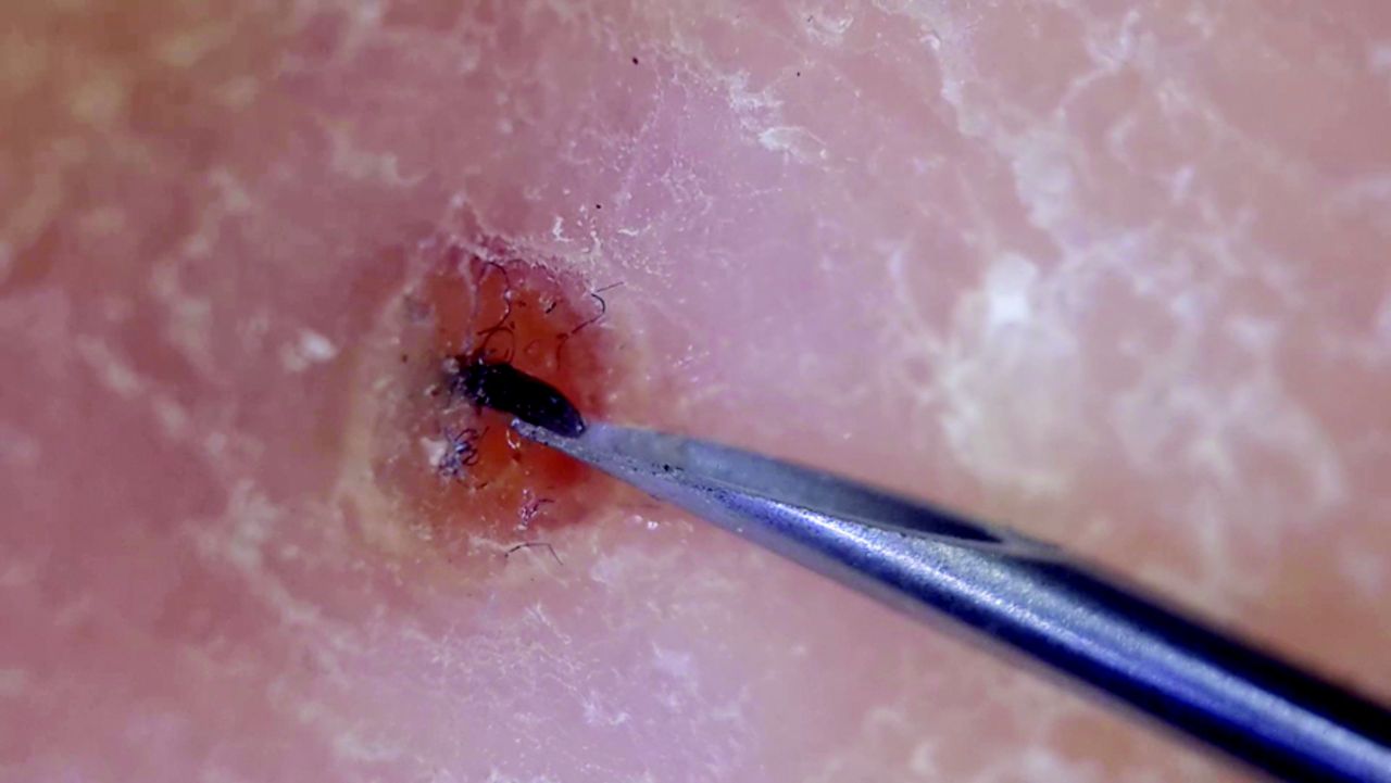
A young man claimed an exquisitely painful heel lasting for the past month. He could not recall walking barefoot outside the household or ever stepping on anything sharp. Even close examination did not reveal the cause of his discomfort. Only a single dark dot on the plantar surface was apparent to the naked eye (Figure 2). Using the dermatoscope, visualization of the protruding tip of a pointed object resulted in the gradual extraction of a long foreign body (Figures 3 and 4). This procedure was completed without any incision. Fortunately, despite the long duration of the indwelling object, piercing perpendicularly deep into the soft tissue, no signs of infection were seen.

Figure 2.
  • Download figure
  • Open in new tab
Figure 2.

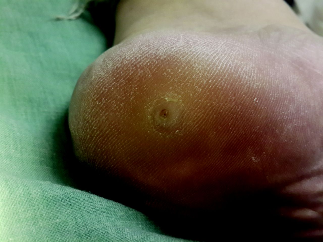
Unassisted appearance of the heel. A subtle break in the skin can be observed.

Figure 3.
  • Download figure
  • Open in new tab
Figure 3.

Juxtamarginal pressure is applied adjacent to tip of the foreign body.

Figure 4.
  • Download figure
  • Open in new tab
Figure 4.

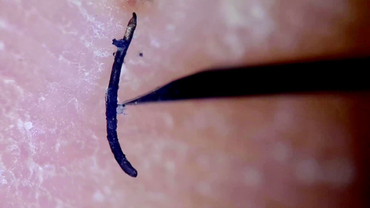
The full size of a rigid, spear-like body upon removal.

Dermoscopy seems to improve the ease and accuracy of locating a penetrating object, the removal of which eventually decreases patients' discomfort. This reduction of symptoms is particularly relevant in specific populations such as young children. The apparent simplicity of the procedure should save time, enabling it to be done during a routine visit.

The use of therapeutic dermoscopy is an acceptable and safe alternative for removing a foreign body. Therapeutic dermoscopy is advocated for use to evaluate and treat many diverse body surfaces, as it increases the likelihood of successful treatment of embedded epidermal objects while decreasing the risk of adverse sequelae from local tissue destruction. Other instances in which this technique has shown its worth for the family physician include delineating the surgical margins of neoplastic skin lesions, revealing ocular pathology, and identifying parasitic infestations.10,13

Notes

  • This article was externally peer reviewed.

  • Funding: none.

  • Conflict of interest: none declared.

  • To see this article online, please go to: http://jabfm.org/content/30/3/374.full.

  • Received for publication April 22, 2016.
  • Revision received December 23, 2016.
  • Accepted for publication January 23, 2017.

References

  1. 1.↵
    1. Eidelman M,
    2. Bialik V,
    3. Miller Y,
    4. Kassis I
    . Plantar puncture wounds in children: analysis of 80 hospitalized patients and late sequelae. Isr Med Assoc J 2003;5:268–71.
    OpenUrlPubMed
  2. 2.↵
    1. Dar TA,
    2. Sultan A,
    3. Hussain S,
    4. Dhar SA,
    5. Ali MF
    . Contracture of the third toe as a delayed presentation of a foreign body in the foot. Foot Ankle Spec 2011;4:298–300.
    OpenUrlCrossRefPubMed
  3. 3.↵
    1. Di Chiacchio N,
    2. Wu SL,
    3. Gabriel FK,
    4. Bet DL,
    5. Di Chiacchio NG
    . Case for diagnosis. Puncture lesions with foreign body in the feet. An Bras Dermatol 2011;86:165–6.
    OpenUrlPubMed
  4. 4.↵
    1. Joseph WS,
    2. Le Frock JL
    . Infections complicating puncture wounds of the foot. J Foot Surg 1987;26 (1 Suppl):S30–3.
    OpenUrlPubMed
  5. 5.↵
    1. Barták V,
    2. Krátký Z,
    3. Popelka ml S
    . Tumour-like lesion of the forefoot due to a retained toothpick fragment. [Case report]. Acta Chir Orthop Traumatol Cech. 2014;81:152–4.
    OpenUrl
  6. 6.↵
    1. Woolfrey PG,
    2. Kirby RL
    . Hypodermic needles in the neuropathic foot of a patient with diabetes. CMAJ 1998;158:765–7.
    OpenUrlAbstract/FREE Full Text
  7. 7.↵
    1. Pattamapaspong N,
    2. Srisuwan T,
    3. Sivasomboon C,
    4. et al
    . Accuracy of radiography, computed tomography and magnetic resonance imaging in diagnosing foreign bodies in the foot. Radiol Med 2013;118:303–10.
    OpenUrlCrossRefPubMed
  8. 8.↵
    1. Nwawka OK,
    2. Kabutey NK,
    3. Locke CM,
    4. Castro-Aragon I,
    5. Kim D
    . Ultrasound-guided needle localization to aid foreign body removal in pediatric patients. J Foot Ankle Surg 2014;53:67–70.
    OpenUrl
  9. 9.↵
    1. Lallas A,
    2. Giacomel J,
    3. Argenziano G,
    4. et al
    . Dermoscopy in General Dermatology: Practical Tips for the Clinician. Br J Dermatol 2014;170:514–26.
    OpenUrl
  10. 10.↵
    1. Naimer SA,
    2. Urkin Y
    . Enhancement of pediatric ophthalmologic diagnosis with hand held polarized dermatoscopy. Clin Pediatr 2014;53:579–84.
    OpenUrlCrossRefPubMed
  11. 11.↵
    1. Naimer SA
    . Prevention and Identification of Retained Sutures. J Am Acad Dermatol 2014;70:e57–8.
    OpenUrl
  12. 12.↵
    1. Naimer SA,
    2. Urkin Y
    . Expanded Use of the Otoscope and Dermatoscope in primary Care. Int J Med & Med Sci 2013;5:535–45.
    OpenUrl
  13. 13.↵
    1. Naimer SA,
    2. Mumcuoglu KY
    . Diagnosis of ophthalmomyiasis externa by dermatoscopy. Dermatol Pract & Conc 2014, 4(4): 85–7.
    OpenUrl
PreviousNext
Back to top

In this issue

The Journal of the American Board of Family     Medicine: 30 (3)
The Journal of the American Board of Family Medicine
Vol. 30, Issue 3
May-June 2017
  • Table of Contents
  • Table of Contents (PDF)
  • Cover (PDF)
  • Index by author
Print
Download PDF
Article Alerts
Sign In to Email Alerts with your Email Address
Email Article

Thank you for your interest in spreading the word on American Board of Family Medicine.

NOTE: We only request your email address so that the person you are recommending the page to knows that you wanted them to see it, and that it is not junk mail. We do not capture any email address.

Enter multiple addresses on separate lines or separate them with commas.
Therapeutic Dermoscopy to Facilitate Detection and Extraction of Foreign Bodies
(Your Name) has sent you a message from American Board of Family Medicine
(Your Name) thought you would like to see the American Board of Family Medicine web site.
CAPTCHA
This question is for testing whether or not you are a human visitor and to prevent automated spam submissions.
10 + 7 =
Solve this simple math problem and enter the result. E.g. for 1+3, enter 4.
Citation Tools
Therapeutic Dermoscopy to Facilitate Detection and Extraction of Foreign Bodies
Sody A. Naimer
The Journal of the American Board of Family Medicine May 2017, 30 (3) 374-376; DOI: 10.3122/jabfm.2017.03.160365

Citation Manager Formats

  • BibTeX
  • Bookends
  • EasyBib
  • EndNote (tagged)
  • EndNote 8 (xml)
  • Medlars
  • Mendeley
  • Papers
  • RefWorks Tagged
  • Ref Manager
  • RIS
  • Zotero
Share
Therapeutic Dermoscopy to Facilitate Detection and Extraction of Foreign Bodies
Sody A. Naimer
The Journal of the American Board of Family Medicine May 2017, 30 (3) 374-376; DOI: 10.3122/jabfm.2017.03.160365
Twitter logo Facebook logo Mendeley logo
  • Tweet Widget
  • Facebook Like
  • Google Plus One

Jump to section

  • Article
    • Abstract
    • Illustrative Case
    • Notes
    • References
  • Figures & Data
  • References
  • Info & Metrics
  • PDF

Related Articles

  • No related articles found.
  • PubMed
  • Google Scholar

Cited By...

  • Research on Clinical Decisions Made Daily in Family Medicine
  • Google Scholar

More in this TOC Section

  • Insurance Instability Among Community-Based Health Center Patients with Diabetes Post-Affordable Care Act Medicaid Expansion
  • Factors Influencing Changing Scopes of Practice Among Contemporary Graduates of the Nation’s Largest Family Medicine Residency
  • Clusters of Health-Related Social Needs Among Adult Primary Care Patients
Show more Brief Report

Similar Articles

Keywords

  • Dermoscopy
  • Foreign Bodies
  • Family Physicians

Navigate

  • Home
  • Current Issue
  • Past Issues

Authors & Reviewers

  • Info For Authors
  • Info For Reviewers
  • Submit A Manuscript/Review

Other Services

  • Get Email Alerts
  • Classifieds
  • Reprints and Permissions

Other Resources

  • Forms
  • Contact Us
  • ABFM News

© 2025 American Board of Family Medicine

Powered by HighWire