
Article Figures & Data
Figures
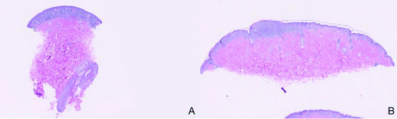
- Figure 1.
Low-power magnification views of biopsied tissue. A: A presumably benign nevus. The section is of a punch biopsy extending into the deep dermis. The lesion in these sections seems to be small, symmetric, and well circumscribed. The melanocytes seem to be well nested, with no upper pagetoid extension, and they mature with descent into the dermis. The melanocytic proliferation extends to the lateral margins, however, so small size, symmetry, and circumscription are not ensured. B: A melanoma. This shave biopsy is a fully adequate specimen because it encompasses the full breadth and depth of the melanocytic proliferation. The proliferation is broad, asymmetric, and poorly circumscribed—characteristics diagnostic of melanoma. In addition, there are areas of randomly dispersed irregular nests of melanocytes, upward pagetoid extension by single melanocytes, and areas of confluence.
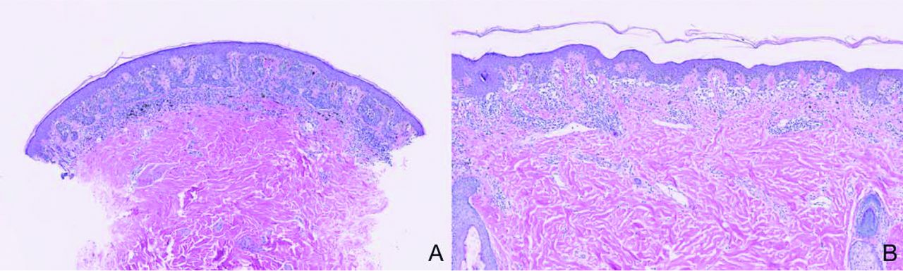
- Figure 2.
High-power magnification views of biopsied tissue. A: A presumably benign nevus. B: A benign-appearing section of melanoma. This area of the melanoma is small, symmetric, and well circumscribed, with small melanocytes that are well nested and located at the base of the rete, with no upper pagetoid extension. A punch biopsy from this area of the melanoma may have rendered an inaccurate benign diagnosis.


Tables
Breadth usually >6 mm Asymmetry Poor circumscription; atypical melanocytes beyond the most peripheral discrete nest of melanocytes within the epidermis Increased number of single atypical melanocytes within the epidermis and epithelial structures of adnexa and, in some foci, single melanocytes predominate over nests of them Melanocytes scattered in the upper reaches of the epidermis Variation in the size and shape of nests of melanocytes Nests of melanocytes are not equidistant Irregular shapes of nests of melanocytes Tendency of nests of melanocytes to confluence Failure of maturation of atypical melanocytes, with progressive descent into the dermis Asymmetrical, patchy distribution of melanin within the neoplasm Extension of atypical melanocytes far down the epithelial structures of the adnexa Asymmetrical distribution of inflammatory cell infiltrates of variable densities at the base of the neoplasm


