
Abstract
Every year, millions of children travel internationally with their families, many to developing countries. Although the vast majority experience uneventful travel and return home well, it is not uncommon for children to present as ill during or after travel. Although the majority of travel-associated illness is mild and self-limited, serious conditions regularly occur. Almost all life-threatening conditions after travel present with fever, and malaria is the most important of these to rapidly exclude. Gastrointestinal symptoms are common after travel in the developing world, and most diarrhea in child travelers has a bacterial source. Children who have a rash in association with fever or who appear ill should receive a priority work-up focused on ruling out serious conditions. Many children traveling internationally experience respiratory illness during or shortly after travel, mainly common upper respiratory infections, yet serious conditions, such as tuberculosis, may occur. Eosinophilia is common in the returned pediatric traveler, particularly those with prolonged stays in the tropics. Not all eosinophilia is caused by parasitic infection; drug reactions, asthma, and other allergic conditions are also common causes. With a focus first on ruling out life-threatening disease and subsequently on an informed and efficient path to diagnosis and treatment, clinicians may confidently provide care for this challenging group of patients.
Every year, millions of children travel internationally with their families, many to developing countries.1 Although the vast majority experience uneventful travel and return home well, it is not uncommon for children to present as ill during or after travel.2 Although the majority of travel-associated illness is mild and self-limited, serious conditions regularly occur.3 Because many health care practitioners in industrialized countries may be unfamiliar with conditions uncommon in their practice setting, this article seeks to review common presentations of the ill child after travel and offer an organized approach to their evaluation.
General Approach
A general approach to the child who presents as ill after international travel is outlined in Table 1. An appreciation of the nuances of common pediatric presentations after travel greatly informs this approach.
Approach to the Child Presenting as Ill after International Travel
Common Presentations
Fever
Almost all life-threatening conditions after travel present with fever.4 Although malaria is the most important of these to rapidly exclude, other potentially serious tropical infections that commonly cause fever after travel include dengue, typhoid, rickettsioses, and leptospirosis; rarely, other serious infections are encountered.2,4
Imported pediatric malaria is not uncommon.5 Children account for 15% to 20% of the several thousand annual cases of imported malaria in industrialized countries,5 and incidence is highest in children who have traveled with caregivers visiting friends and relatives (travelers known as VFRs).5 Although Plasmodium falciparum is the most dangerous malarial strain, any malaria in a nonimmune child (most travelers) is serious6; malaria caused by Plasmodium vivax, for example, may also be associated with severe malaria.7,8 Particularly in infants and smaller children, malaria may present with protean manifestations, including predominantly gastrointestinal symptoms such as abdominal pain or vomiting, making diagnosis challenging unless a high index of suspicion exists.6
Whether or not the child received antimalarial chemoprophylaxis is an important historic detail, but of greatest importance is whether or not the child was exposed to malaria; malaria may develop even when prophylaxis has been properly taken.6 Any ill, febrile child who has traveled in a malaria-endemic zone (see map at www.cdc.gov/malaria/risk_map/) in the year before presentation could have malaria.9 The risk of severe malaria is higher for those who present during the first month after travel because P. falciparum's incubation period is typically 10 to 30 days (range, 7–90 days),4 and for those who have traveled to sub-Saharan Africa (where P. falciparum is the most common malarial strain).5
Incubation periods (Table 2) give critical clues to the etiology of fever after travel.1 For example, in much of the tropics, areas endemic for malaria and dengue overlap. Fever that develops within 7 days of entering an endemic area should not be caused by malaria, whereas fever developing more than 2 weeks after leaving an endemic area should not be because of dengue.10
Incubation Periods for Selected Infections Responsible for Fever in Children Who Have Returned from the Tropics
Initial investigations for a febrile child without a localizing source of infection vary based on the individual history, but will probably include a complete blood count, liver function tests, urinalysis, blood culture, and peripheral blood smears for malaria.11,12 Additional testing may include cultures of stool and urine, chest radiography, and specific serologic assays such as those for dengue, rickettsiae, schistosomes, and Leptospira.11,12
The proper management of malaria depends on the diagnosis being swift and reliable. Alternatives for malaria diagnosis exist. Traditionally, malaria has been diagnosed with blood smears; a minimum of 3 sets must be performed before malaria can be tentatively ruled out in a child who has a history of possible malaria exposure and clinical signs and symptoms potentially consistent with the disease.6 Yet, although serial blood smears have very high specificity,13 their sensitivity is relatively low14 and quite dependent on the experience level of their interpreter, which may be limited in many nonendemic settings.9 Rapid diagnostic tests (RDTs) based on the detection of species-specific, histidine-rich proteins or lactate dehydrogenase exist and will readily both diagnose malaria and differentiate between falciparum and nonfalciparum infections.15,16 In field and clinical trials, several of these compared well with blood smear analysis in the detection of P. falciparum.15,16 One recently reported randomized clinical trial demonstrated 95.4% sensitivity and 95.9% specificity in the detection of malaria using RDTs.16 Molecular diagnostics (such as polymerase chain reaction) are particularly useful where malaria incidence is low and false-negative smears or RDT results are more likely to be accepted as valid (as in most industrialized settings where children ill after travel are evaluated).17 Even when initial malarial tests are negative, children who have a history of malaria exposure and suggestive symptoms should be approached as if they have malaria until an alternative diagnosis is made.6,12
Specific management of malaria depends on whether the case is severe or uncomplicated and on the particular or suspected malarial strain (particularly whether or not the strain is P. falciparum).5,9,18,19 Most countries have guidelines for malaria management,9,19 as does the World Health Organization.18 Unless a practitioner has experience with malaria diagnosis and management, early consultation with an infectious disease or tropical medicine specialist is advisable, and most national authorities have a service comparable to the Centers for Disease Control and Prevention in the United States, which operates a Malaria Hotline for assistance with suspected or confirmed malaria cases (1-770-488-7788).
A major public health threat in the tropics, dengue has become the most common diagnosis in febrile travelers returned from all tropical regions except Africa.10 Dengue has a short incubation period and generally results in self-limited illness, although as many as 250,000 cases of severe dengue (hemorrhagic and shock syndrome forms) occur annually, including cases in returned travelers.10 Dengue risk for travelers is sometimes characterized as the “opposite” of that for malaria: most commonly transmitted in urban areas during the daytime (where and when its principal vector, Aedes aegypti, lives and feeds) whereas malaria risks are generally higher from dusk to dawn (when its Anopheles spp. vector feeds) and in rural areas.20
A clinical diagnosis, dengue presents after exposure in an endemic area with fever, uncomfortable constitutional symptoms and, often, a characteristic maculopapular rash (Figure 1), which may be less apparent in children who have darker complexions.21 Leukopenia and thrombocytopenia are common laboratory findings, and useful serologies and rapid diagnostic tests may be available in some settings.22

Maculopapular rash with diffuse petechiae and areas of bruising associated with dengue hemorrhagic fever. Photo courtesy of the Emerging Infectious Diseases journal. Reprinted from http://www.cdc.gov/EID/content/14/8/1329-G.htm.
Care of Patients Varies by Degree of Dengue Severity
Dengue hemorrhagic or shock syndrome is rare with primary dengue infection (the case with most travelers).10,21 Nonsevere dengue is self-limited and care is supportive, whereas severe dengue requires hospitalization and intensive management focused on early recognition and treatment of shock.21 Development of severe dengue is heralded by hemoconcentration (increasing hemoglobin); thickening of the gallbladder wall has been shown to predict the development of vascular permeability (the hallmark of severe dengue21) in children who have dengue.23
The Chikungunya virus (also transmitted by Aedes spp mosquitoes) overlaps epidemiologically with dengue and causes a febrile arthralgia syndrome. Millions of cases of chikungunya have been seen in Africa, the Indian Ocean basin, the Indian subcontinent, and Southeast Asia since 2005, including thousands in returned travelers.24 The incubation period is similar to dengue and a similar rash may be present, but severe disease is rare, particularly in children.25 Treatment is symptomatic, and illness caused by chikungunya is usually self-limited but substantial joint pain may persist.26
Yellow fever is a serious arboviral disease with a high fatality rate, yet is very rare in travelers because of the availability of an effective vaccine.27 Other arboviral illnesses may occur, but are rarely reported in travelers.22
Virtually indistinguishable clinically,11 typhoid and paratyphoid (enteric fever) are caused by fecal-oral transmission of Salmonella typhi and Salmonella paratyphi, respectively. Risk is higher for VFRs than for traditional tourists and for those who have traveled to the Indian subcontinent, where incidence is highest.12 Symptoms include fever and abdominal pain, myalgias and arthralgias, nausea and vomiting, and occasionally diarrhea.28 Some patients may exhibit a characteristic, if uncommon, “stepladder” fever pattern, with febrile temperature progressively increasing over time.28 Relative bradycardia is a classic associated sign (although also seen with other tropical infections), as are splenomegaly, rose spots, and, occasionally, confusion.28
Laboratory studies often show a normal or decreased white blood cell count, and liver enzymes are usually elevated. Diagnosis of typhoid occurs via culture. Bone marrow cultures are most sensitive (80% to 95%), whereas blood cultures and stool cultures show variable sensitivity (blood cultures are 70% sensitive during the first week and 30% sensitive during the second week; stool cultures are more sensitive as the disease progresses3).
Although 10% to 15% of patients will have complications, including life-threatening gastrointestinal bleeding or perforation, complications are more frequent in younger patients and those who have been ill more than 14 days.12
Antibiotic treatment depends on the region of the world where infection was acquired, with Latin American and Caribbean strains presumed to be fully sensitive (amoxicillin, trimethoprim-sulfamethoxazole, quinolones); South Asian strains may be multidrug resistant, including to quinolones, requiring azithromycin or cefixime.12,28–30 Severe illness should, of course, be treated in a hospital with parenteral antibiotics.
Rickettsial infections should be considered in the differential diagnosis of the febrile returned child, particularly African tick-bite fever, caused by Rickettsia africae and increasingly common in recent years, particularly in southern Africa, including popular game parks.11 African tick-bite fever classically presents with a primary eschar at the site of the tick bite, followed by a flu-like illness with generalized rash.11 Diagnosis of rickettsial illness can be made by specific serology; treatment for adults and children is with doxycycline.31
Not restricted to the tropics, leptospirosis acquisition by travelers usually occurs through recreational exposure to water or soil contaminated with infected animal urine, with outbreaks more common after heavy rainfall.12 Leptospirosis should be considered in the febrile returned child who is systemically ill and who has tested negative for malaria, particularly if characteristic features such as conjunctival inflammation are present. Although serologic diagnosis is available, it may be of low yield.12 Severe leptospirosis may progress to jaundice, renal failure, and hemorrhage, and case fatality rates can be high.12 Children should be treated with ampicillin or doxycycline, and severe disease should be treated with intravenous ceftriaxone or penicillin G.12
Gastrointestinal Symptoms
Although generally not possessing the serious nature that fever may, gastrointestinal symptoms are nonetheless common after travel in the developing world.
Most traveler's diarrhea (TD) in children is caused by bacteria, but in children younger than age 2, viral etiologies (norovirus, rotavirus if not immunized) may be more common, as may be atypical and/or prolonged episodes of TD.32,33 TD is usually self-limited and often warrants simple observation. Azithromycin is the treatment of choice for most bacterial etiologies; 10 mg/kg once daily for 3 days is a common regimen.34 Helpful laboratory studies include stool cultures, giardia antigen, hemoccult, and fecal leukocytes; when red and white blood cells are both absent from the stool, the negative predictive value to exclude invasive organisms exceeds 95%.3
Persistent diarrhea (present >2 weeks) is more likely to have a protozoal source (such as Giardia lamblia or Cryptosporidium).32 Stool samples for ova and parasite analysis should be performed to exclude parasitic sources. Even a single sample has utility; data from studies of serial stool sample analysis show >90% of parasites are detected in the first sample submitted.35 Testing for Giardia antigen can increase the yield in the diagnosis of this common parasitic cause of persistent diarrhea and may need to be tested repeatedly.36 Likewise, special stains of stool for Cryptosporidium and Cyclospora are often necessary to identify these organisms, which in some series have been shown to be relatively common causes of persistent diarrhea in returned travelers.37 When there is a history of antibiotic exposure, testing for Clostridium difficile may yield a diagnosis.38 As mentioned above, infants and young children may experience prolonged episodes of diarrhea when infected with bacterial organisms that commonly cause acute, self-limited diarrhea in older individuals; for this age group, stool cultures may be useful in the evaluation of persistent diarrhea.32
When diarrhea persists and repeated investigations for infection are negative, noninfectious causes, such as secondary lactose intolerance after infection, should be considered. Irritable bowel syndrome after infection is also common after an episode of TD, as is the “unmasking” of autoimmune diseases, such as inflammatory bowel disease or celiac sprue.33
Tropical sprue is a rare cause of persistent diarrhea that has malabsorptive features; most commonly seen after travel to South Asia, its incidence is declining.33 It generally responds to a course of broad-spectrum antibiotics.39 Helminthic infections (roundworms) are unlikely in traveling children, but may be more common in children who have had prolonged stays in the tropics or children traveling with VFRs; with heavy infestations there may be symptoms.3
Uncommonly, children may also present with jaundice after travel in the developing world, and this may herald serious illness.3 Febrile jaundice should prompt a brisk and thorough assessment for life-threatening disease.3 Although viral hepatitis (primarily caused by hepatitis A) can also present with fever, symptomatic hepatitis A infection in traveling children is uncommon.40 Hepatitis E is a common cause of acute hepatitis in the developing world, shares a common fecal-oral route of transmission with hepatitis A, and should be considered when hepatitis A testing is negative.41
Dermatologic Symptoms
Skin conditions are common after travel to the tropics, and in large, retrospective reviews have been noted to occur disproportionately after travel to the Caribbean or Central or South America.2,42 As described above, many tropical infections, such as dengue, present with skin findings, as do many cosmopolitan infections, such as measles, meningococcemia, and rickettsioses. Rash—particularly if petechial or hemorrhagic—in association with fever or an child who appears ill should receive a priority workup focused on ruling out serious conditions.11,12,21
Fortunately, serious underlying disease is rare in children who present with skin conditions after travel.42 In a review of infants to children aged 17 years who presented to a large, global network of tropical and travel medicine clinics (GeoSentinel), the 5 most common skin conditions after travel were cutaneous larva migrans, dog bites, insect bites (including superinfected), cutaneous leishmaniasis, and superficial skin abscesses.42
Diagnosis of common tropical skin conditions tends to be straightforward; clinical diagnoses, aided by knowledge of geographical disease trends, are the rule. Dog bites, insect bites, and superficial skin abscesses are clear diagnoses, whereas urticaria suggests allergic or parasitic exposure, with schistosomiasis and strongyloides being common causes of urticaria among the latter group.3
Cutaneous leishmaniasis in travelers most commonly presents several weeks after travel to Central and South America as an ulcerated skin lesion (having originated as a papulonodular lesion at the site of a sand fly bite) surrounded by a raised, indurated border42,43; it is also a common condition in the Middle East, Iran, and Afghanistan.43
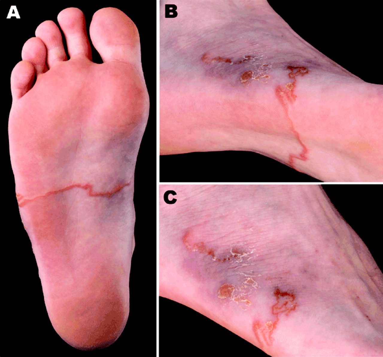
Cutaneous larva migrans occurs after exposure of bare skin to sand, and, not surprisingly, tends to follow tropical beach holidays.44 An intensely itchy, migratory rash (Figure 2), usually around the foot or ankle, but possible anywhere skin contacted sand (such as the back), represents subcutaneous movement of the larval stage of the dog hookworm.44

Cutaneous larva migrans. A and B: Typical elevated lesion caused by the migrating parasite on the plantar surface of the foot. C: Ulcerative lesion at site of origin on the lateral side of the foot. Photo courtesy of the Emerging Infectious Diseases journal. Reprinted from http://www.cdc.gov/eid/content/15/11/1856-F.htm.
Myiasis is another common skin condition imported from the tropics, occurring when a fly larva infests skin, creating a painful, boil-like lesion. Tumbu fly myiasis is seen in travelers returning from tropical Africa whose clothing was contaminated with fly eggs, usually while hanging on a washing line; ironing clothes with a hot iron after line drying may be preventive.45 Bot fly myiasis presents in travelers returning from Central and South America; unlike the Tumbu fly, the bot fly deposits its eggs directly on the skin.45 Management of myiasis and other skin conditions presenting commonly after tropical travel is detailed in Table 3.
Management of Common Skin Conditions Presenting after Travel to Developing Countries
Respiratory Symptoms
As many as 25% of children traveling internationally may experience a respiratory illness during or shortly after travel, mainly common upper respiratory infections such as pharyngitis, sinusitis, and otitis media; air travel alone is a risk factor for these conditions.1 The child who presents with flu-like illness outside of influenza season could still have influenza because influenza circulates year-round in the tropics, and in “summer” (by “home country” definition) in the opposite hemisphere. Although exotic pulmonary infections are reasonably uncommon in travelers, travel histories and regional disease trends are very important when respiratory symptoms are present, as is demonstrated by reports of acute histoplasmosis (headache, fever, cough, myalgias) in a large group of children who were visiting a cave site in Costa Rica47; melioidosis (usually pneumonia) in returned travelers from Southeast Asia and northern Australia48; and, most recently, H1N1 influenza in children who had recently visited Mexico.49
Active pulmonary tuberculosis (TB) in returning pediatric travelers is rare, and, if present, should be associated with chest film findings.3 All children who have potentially been exposed to TB during travel (known exposure; prolonged travel in developing countries; and, in some settings, travel with VFRs3) should be screened for latent TB with placement of purified protein derivative. This is particularly important for infants and young children because severe forms of TB are more common in children younger than 5 years of age (although rates are lower in those who have received Bacille Calmette-Guérin immunization).
Eosinophilia
Eosinophilia (absolute eosinophil count >500/uL) is common in the returned pediatric traveler, particularly those who stayed in the tropics longer than 3 months (10%).3 Not all eosinophilia is caused by parasitic infection; drug reactions, asthma, and other allergic conditions are also common causes, although high absolute eosinophil counts (>1,000) in the returned traveler are predictive of parasitic infection.3
Schistosomiasis is a particularly common parasitic cause of eosinophilia after exposure to fresh water in the tropics.50 Although acute schistosomiasis (Katyama fever) is a cause of fever and urticaria accompanying eosinophilia, schistosomiasis is more commonly discovered in the returned pediatric traveler as positive serology during the workup of asymptomatic eosinophilia noted some time after return or on request by caregivers for a test after known exposure.51 Other common parasitic causes of eosinophilia include geohelminths, particularly hookworm and strongyloides, and filariasis3; these are more likely in children returning from travel with VFRs and children emigrating or presenting as refugees from endemic tropical regions.50,51 An approach to the evaluation and management of eosinophilia after travel is detailed in Table 4.
Evaluation of Eosinophilia Presenting after Travel to Developing Countries
Conclusion
Most children travel to developing countries and return home without incident. However, although serious illness during or after travel is uncommon, presentation to a clinician with a health-related concern after travel is not. In the evaluation and treatment of a child presenting with such concerns, clinicians must entertain diagnostic possibilities that would not ordinarily be considered had the child not traveled. With a focus first on ruling out life-threatening disease, such as malaria, and subsequently on an informed and efficient path to diagnosis and treatment, clinicians may confidently provide care for this challenging group of patients. Table 5 reviews Strength of Recommendation suggestions for evaluation and treatment of the child who presents as ill after international travel.
SORT Recommendations54 for the Evaluation and Treatment of a Child Who Presents as Ill after International Travel
Notes
This article was externally peer reviewed.
Funding: none.
Conflict of interest: none declared.
- Received for publication December 4, 2009.
- Revision received June 8, 2010.
- Accepted for publication June 14, 2010.


