Figure 1.
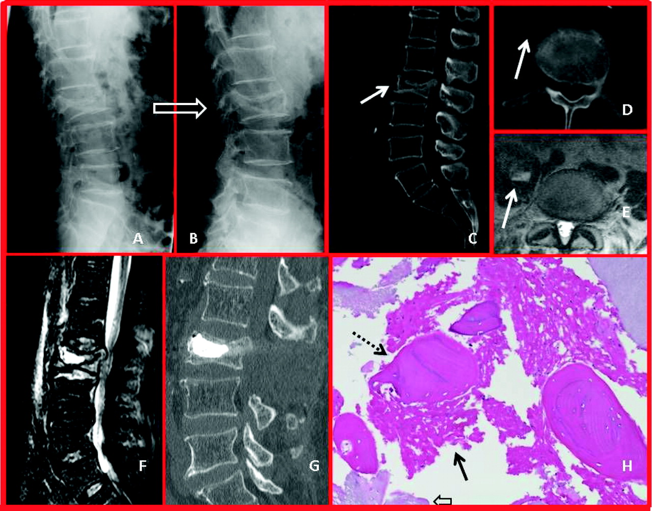
The following sequence of images illustrates the salient radiologic and pathologic findings in Kummel disease. In flexion, the fracture compresses through the avascular zone directly beneath the superior endplate, demonstrating the maximal degree of height loss, but in extension, the fracture plane gaps open (A and B). This motion (pseudarthrosis) is responsible for the vacuum effect, which pulls transudative fluid and nitrogen gas (white arrow, C) into the fracture site. Intervertebral gas is a radiographic and computed tomographic finding indicative of Kummel disease. In this case, the fluid and gas escaped from the fracture and descended along the right psoas muscles, creating the false appearance of a psoas abscess (D). Confirming this explanation of the fluid collection over the right psoas, the fluid collection is benign in appearance; there is no associated soft-tissue edema on T2 magnetic resonance images (MRIs) (E). Sagittal STIR MRI, the image modality of choice for diagnosing acute VCFs, shows fluid (homogeneously bright white area) in the fracture cleft, with air bubbles (black dots). Fluid filled clefts on MRIs are characteristic of Kummel disease and occur because patients undergoing MRI lie supine for ≥40 minutes, which seems to be enough time to allow transudative fluid to replace intervertebral gas. Note, the vertebral body height is greater on the MRI (F) than on the flexion radiograph (A), and this is because the supine position extends the spine. The most important finding on the MRI is that the adjacent level endplates and vertebral bodies (L2 and L4) are completely normal. Spondylodiscitis or vertebral osteomyelitis would have destroyed and/or inflamed the adjacent disk spaces and endplates. The pathopneumonic finding in this sequence of images, is the intervertebral gas, pseudarthrosis and lack of adjacent inflammation. The postoperative sagittal CT-recon image shows the results of open kyphoplasty, with laminectomy (note that the L3 spinous process is absent) and improvement in L3 vertebral height (G). H: The final image is a high power view (original magnification, 100×) of the biopsy obtained at surgery, which shows areas of necrotic bone (dotted arrow); immature, woven bone (solid arrow); and nests of cartilage, all which are pathopneumonic for Kummel disease.


