
There have been rare case reports in the specialty medical literature describing an association between Epstein-Barr virus (EBV) infection and genital ulcerations in women and men.1–6 This association is not well known to primary care physicians, however, and it is not described in various widely used reference texts.7–9 Lack of awareness of this condition can lead to expensive, unnecessary diagnostic testing; misdiagnosis as herpes simplex virus (HSV) infection or even possible sexual abuse has serious consequences for the patient. Here, we present 2 case reports of infectious mononucleosis in which severe genital ulceration was the presenting symptom.
Case Reports
Patient A was an 18-year-old woman who presented with multiple hemorrhagic, extremely tender, raised lesions on the labia minora. Sexual history involved oral and manual contact with one female partner in her lifetime; the most recent episode was 2 weeks before presentation. She had complained of a mild sore throat and fatigue 1 week prior, although her throat appeared normal at the time of presentation. The lesions evolved into 1-cm exudative ulcerations associated with fever. The pain was so severe that the patient required narcotic analgesia and had great difficulty walking. She was treated with oral acyclovir for presumed herpes simplex virus infection. One week after the onset of the vulvar lesions, the patient developed an exudative tonsillitis and cervical lymphadenopathy. The Monospot test for heterophile antibodies and complete blood count performed at this time were consistent with acute infectious mononucleosis. Results of other laboratory tests, including herpes simplex virus culture, gonorrhea, chlamydia DNA probe testing, and throat culture for group A β-hemolytic streptococci, were all negative. Bacterial culture obtained from the lesions grew normal flora. The vulvar ulceration healed completely without scarring after approximately 18 days.
Patient B was an 18-year-old woman who presented with multiple painful, hemorrhagic lesions of the inner labia minora. She had no history of any prior sexual activity, but because the lesions raised the suspicion of herpes simplex virus infection, acyclovir was prescribed.
Over the course of the subsequent 2 to 3 days, the sores coalesced into large, extremely painful necrotic areas from which there was significant sloughing of tissue. Fever spiked briefly on day 3. A complete blood count on day 5 showed a white cell count of 5000/dL with 24% atypical lymphocytes. Platelets were slightly reduced at 131,000. This suggested infectious mononucleosis, and examination showed early tonsillar exudates and minimal cervical lymphadenopathy. A Monospot test for heterophile antibody was negative, however, as were tests for Epstein-Barr viral capsid antigen IgG and IgM antibodies. The patient was prescribed ciprofloxacin pending a Haemophilus ducreyi culture result, which later proved negative, as were a direct fluorescent antibody test for HSV, HSV type-specific serology, and rapid plasma reagin testing. Bacterial culture from the lesions resulted in the growth of normal flora.
By day 8, the patient had developed the typical clinical picture of infectious mononucleosis, with severe exudative tonsillitis and anterior and posterior cervical lymphadenopathy. The Monospot became weakly positive on day 10 and definitively positive on day 15; serum drawn that day was positive for both IgG and IgM antibodies to Epstein-Barr viral capsid antigen. Throat culture for group A β-hemolytic streptococci was negative.
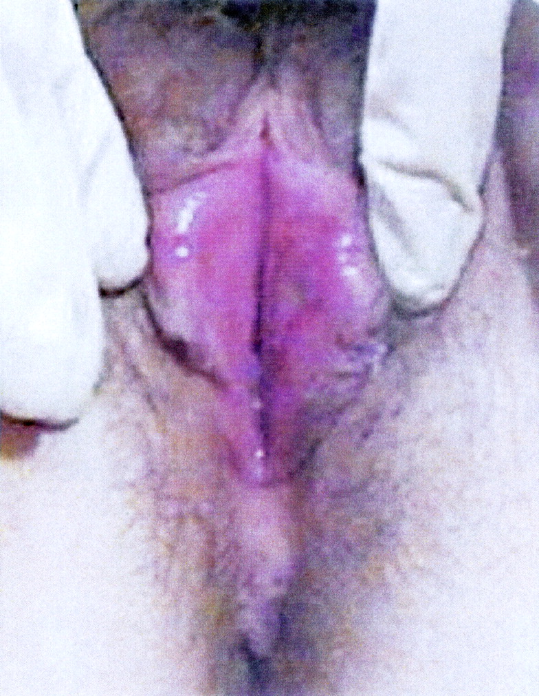
Once the diagnosis of infectious mononucleosis was clear, acyclovir and ciprofloxacin were discontinued, and the patient was treated with prednisone because of very severe tonsillar swelling. The ulcers healed gradually over the following week (Figure 1), with resolution of all symptoms by day 22.

Patient B. Healing ulceration on day 18.
Epstein-Barr viral capsid antigen IgM antibody is normally detectable before or around the time of the appearance of symptoms of infectious mononucleosis. It is notable that in this patient, neither heterophile nor specific antibodies were detected, despite the presence of severe genital ulceration, until later in the course of the illness, when typical mononucleosis symptoms had also developed.
Discussion
These two cases are of particular interest because of the severity of the ulcerations, the lack of other clinical or laboratory evidence of acute EBV infection at the time of presentation, and the absence of any possibility of sexual transmission in patient B. Other diagnoses that we considered in these patients included HSV, syphilitic chancre, chancroid, bacterial infection, lichen planus or Behcet disease, all of which were ruled out based on either clinical presentation or negative lab testing. Although we did not obtain EBV cultures directly from the ulcerations, it seems highly likely given the clinical and laboratory picture and absence of other cause that EBV was the underlying cause of the ulcerations. Direct isolation of EBV from a genital ulceration has been reported,10 and other studies have confirmed the presence of EBV in the cervix,11,12 lending support to this hypothesis. Several of the case reports cited in the specialty medical literature involved women who had never been sexually active,1–5 similar to patient B, suggesting either autoinoculation of the genitals and/or hematogenous spread.
This condition may be more common than is recognized and may often be mistakenly treated as HSV. Increased awareness by clinicians of EBV as a possible cause of genital ulcers will lead to improved patient care.
- Received for publication October 11, 2004.
- Revision received October 11, 2004.


Related Articles
Cited By...
- No citing articles found.