
Injuries to the tarsometatarsal joint, also known as Lisfranc injuries, are uncommon and can be difficult to diagnose. These injuries have a high potential for causing substantial disability related to posttraumatic osteoarthritis. As a result, it is important to be alert for these injuries when evaluating patients after acute foot trauma. This case report describes a collegiate athlete with a Lisfranc fracture-dislocation. Relevant aspects of the physical examination, diagnostic imaging, and treatment considerations are discussed.
Case Report
The patient, a 22 year-old male collegiate football player, was examined on the field after sustaining an injury to his right foot. After falling on a plantar-flexed, fixed foot, an axial load was applied to the posterior aspect of the patient’s right heel. He developed immediate pain and swelling over the dorsum of the foot, and he was unable to bear weight on his foot. His medical and surgical history were unremarkable.
When the patient was examined, he was in extreme discomfort. He had considerable swelling over the dorsum of the midfoot. His foot was diffusely tender with areas of maximal tenderness over the first and second tarsometatarsal joints, the medial cuneiform, and between the first and second metatarsal heads. The foot was otherwise neurologically intact.
The patient was sent to the local emergency department for radiographs immediately after the injury. Non-weight-bearing anteroposterior, lateral, and oblique radiographs were obtained. The physicians caring for the patient in the emergency department, including the on-call radiologist, did not observe any abnormalities on the initial films. The patient was released from the emergency department with crutches and a postoperative shoe. He was told that he had a severe sprain of his foot. When the patient brought the radiographs to the training room the next day, a fracture of the medial cuneiform was seen in addition to 2 mm of widening between the medial and middle cuneiform articulation, findings consistent with a Lisfranc fracture-dislocation (Figure 1).
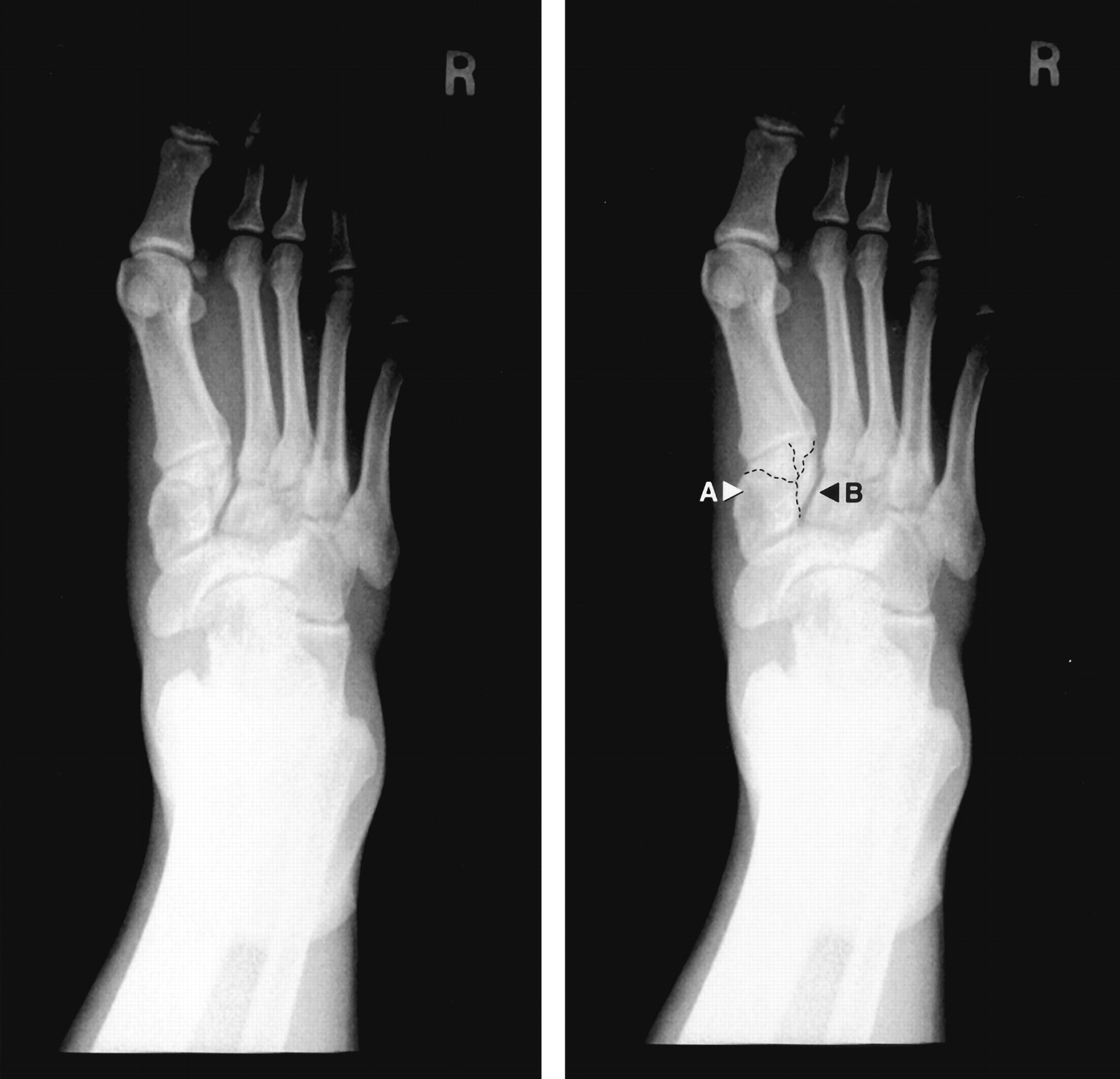
A 22-year-old man with midfoot pain and swelling. Panel 1: weight-bearing anteroposterior radiograph of the right foot. Panel 2: medial cuneiform is labeled (A), and fractures of the medial cuneiform are defined (dotted lines). A 2-mm diastasis is noted between the medial and middle cuneiforms (B).
The patient’s foot was placed in a fracture boot, and he was assigned to strict non-weight-bearing status. He was then referred to a foot and ankle surgeon, who took him to the operating room 4 days after the injury for open reduction and internal fixation of both the medial cuneiform fracture and the Lisfranc dislocation. A cannulated screw was placed from the medial aspect of the medial cuneiform across the Lisfranc articulation into the base of the second metatarsal. A second screw was placed across the medial cuneiform into the middle cuneiform (Figure 2). The patient was instructed to remain non-weight bearing, and his foot was put in a cast to immobilize it. He was then given a fracture boot to wear for 6 weeks. He was gradually weaned from his fracture boot without difficulty. The hardware was removed 19 weeks after surgery. At 42 weeks after surgery and 22 weeks after removal of the screws, the patient was asymptomatic and was given permission to participate in all activities without restrictions. He participated in preseason conditioning without incident and was planning to compete in the regular season.
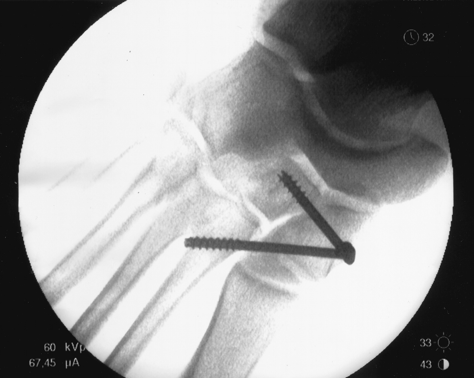
Intraoperative film showing cannulated screw placed from the medial aspect of the medial cuneiform across the Lisfranc articulation into the base of the second metatarsal. A second screw is placed across the medial cuneiform into the middle cuneiform.
Discussion
The Lisfranc joint complex is composed of the articulations between the tarsal and metatarsal bones. Transverse ligaments join the bases of all the metatarsals with the exception of the articulation between the first and second metatarsals.1 ,2 The Lisfranc ligament spans the medial cuneiform and the base of the second metatarsal. The base of the second metatarsal lies within a mortise created by the three surrounding cuneiform bones. This keystone arrangement confers stability to the joint despite the absence of a ligamentous connection between the first and second metatarsal heads. There is little support provided to the dorsal surface of the tarsometatarsal joint.3 ,4 As a result of this configuration, when the foot is positioned in extreme plantar-flexion, the second metatarsal is prone to dislocate dorsally if an axial load is applied. Because of the keystone configuration of the base of the second metatarsal and the strength of the Lisfranc ligament, a fracture at the base of the second metatarsal is commonly observed with a Lisfranc dislocation.
Injuries to the Lisfranc region are uncommon and are classically described in the trauma literature.2 They occur most commonly as a result of a motor vehicle accident.4 Rarely have they been described as a result of participation in sports activities. The annual incidence is estimated to be 1 in 55,000 persons per year.5 ,6 It is important to diagnose these injuries early and initiate appropriate treatment. The most common long-term complication of a Lisfranc joint injury is chronic pain secondary to posttraumatic osteoarthritis, particularly if the congruency and stability of the Lisfranc articulation are not reestablished.2 ,3 ,5 ,7–9
Lisfranc joint injuries can be difficult to recognize and are commonly misdiagnosed during the initial examination by a health care provider.3 ,10 ,11 Midfoot swelling and the inability of the patient to bear weight on the affected foot, either immediately after the injury or when examined in the office, might be the only clues that this injury has occurred.3 The physical examination should focus on eliciting tenderness over the tarsometatarsal articulations. In addition, stress should be applied to the tarsometatarsal articulations by passive supination and pronation of the forefoot. This test could be the only physical examination maneuver that reproduces discomfort in subtle injuries to this area. It is important to eliminate subtalar motion by maintaining the hindfoot in a position of inversion before performing this test, as a notable amount of supination and pronation of the foot occurs at the subtalar joint.
For patients who complain of foot pain after a classic mechanism of injury and the above findings are observed on physical examination, a strong suspicion of a Lisfranc fracture-dislocation should prompt the clinician to obtain further imaging studies. The physician should also continue to look for this fracture-dislocation when interpreting radiographs, as it has been estimated that 20% of Lisfranc joint injuries are missed on initial radiography.2 ,4 ,12 These studies should include weight-bearing anteroposterior, lateral, and oblique radiographs of the foot because non-weight-bearing views of the foot can be normal.2 ,3 Foster and Foster13 showed that the most consistent radiographic finding in Lisfranc joint injuries was the loss of alignment of the medial border of the second metatarsal and the medial border of the middle cuneiform. If a Lisfranc injury is suspected but not confirmed by radiographs, magnetic resonance imaging or computed tomography of the foot should be considered.5
Once the diagnosis is established, the optimal treatment approach and prognosis are subject to controversy. Even though there is little consensus regarding the correlation of long-term outcome to the degree of diastasis between the first two metatarsal heads, current management options are based on the concept that more satisfactory results will ensue from a stable, anatomic reduction of the fracture-dislocation.2 ,4 ,12 Most investigators have concluded that there is little place for the nonoperative management of Lisfranc fracture-dislocations when a 2-mm or greater diastasis is found between the bases of the first and second metatarsals and medial and middle cuneiforms, because it is difficult to maintain anatomic reduction by closed reduction and immobilization alone.1 ,2 ,4 ,5 ,7 ,9 ,14 ,15
Curtis et al16 recommend surgical reduction for all athletes and active persons. Surgical reduction as soon as possible after the injury is recommended by most orthopaedic surgeons. Trevino and Kodros12 recommend surgical reduction within the first 24 hours after severe injuries. A delay of 1 to 2 weeks, however, might be appropriate to allow resorption of soft-tissue swelling and does not appear to alter long-term outcomes.5 Surgical correction after 6 weeks generally results in poor functional outcomes.12
Closed or open reduction with Kirschner wire (K wire) internal fixation has historically been used for the treatment of these injuries.14 The use of open reduction and internal fixation with AO screws (meets Arbeitsgemeinschaft für Osteosynthesefragen international standards) has grown increasingly popular for Lisfranc fracture-dislocations.12 After screw fixation, most orthopedists recommend immobilization and non-weight-bearing status for 8 to 12 weeks. The screws may be removed at 12 weeks. Full weight bearing is typically not permitted until all hardware is removed.
Conclusion
It is essential to be highly aware of a possible Lisfranc fracture-dislocation when evaluating patients who have midfoot pain and swelling. Liberal use of weight-bearing radiographs is appropriate when a Lisfranc fracture-dislocation is suspected. Appropriate management, including early surgical consultation, is required to improve the long-term functional outcome.
- Received for publication September 10, 2002.
- Revision received September 10, 2002.


