
Main menu

  • HOME
  • ARTICLES
    • Current Issue
    • Ahead of Print
    • Archives
    • Abstracts In Press
    • Special Issue Archive
    • Subject Collections
  • INFO FOR
    • Authors
    • Reviewers
    • Call For Papers
    • Subscribers
    • Advertisers
  • SUBMIT
    • Manuscript
    • Peer Review
  • ABOUT
    • The JABFM
    • The Editing Fellowship
    • Editorial Board
    • Indexing
    • Editors' Blog
  • CLASSIFIEDS
  • Other Publications
    • abfm

User menu

Search

  • Advanced search
American Board of Family Medicine
  • Other Publications
    • abfm
American Board of Family Medicine

American Board of Family Medicine

Advanced Search

  • HOME
  • ARTICLES
    • Current Issue
    • Ahead of Print
    • Archives
    • Abstracts In Press
    • Special Issue Archive
    • Subject Collections
  • INFO FOR
    • Authors
    • Reviewers
    • Call For Papers
    • Subscribers
    • Advertisers
  • SUBMIT
    • Manuscript
    • Peer Review
  • ABOUT
    • The JABFM
    • The Editing Fellowship
    • Editorial Board
    • Indexing
    • Editors' Blog
  • CLASSIFIEDS
  • JABFM on Bluesky
  • JABFM On Facebook
  • JABFM On Twitter
  • JABFM On YouTube
Review ArticleClinical Review

Diagnosis and Management of Atopic Dermatitis for Primary Care Providers

Patrick Fleming, Yue Bo Yang, Charles Lynde, Braden O'Neill and Kyle O. Lee
The Journal of the American Board of Family Medicine July 2020, 33 (4) 626-635; DOI: https://doi.org/10.3122/jabfm.2020.04.190449
Patrick Fleming
From Division of Dermatology, University of Toronto, Toronto, ON, Canada (PF, CL); MD Program, University of British Columbia, Vancouver, BC, Canada (YBY); Department of Family and Community Medicine, North York General Hospital, Toronto, ON, Canada (BO); Department of Family and Community Medicine, St. Michael's Hospital, Toronto, ON, Canada (KOL).
MD, MSc, FRCPC
  • Find this author on Google Scholar
  • Find this author on PubMed
  • Search for this author on this site
Yue Bo Yang
From Division of Dermatology, University of Toronto, Toronto, ON, Canada (PF, CL); MD Program, University of British Columbia, Vancouver, BC, Canada (YBY); Department of Family and Community Medicine, North York General Hospital, Toronto, ON, Canada (BO); Department of Family and Community Medicine, St. Michael's Hospital, Toronto, ON, Canada (KOL).
BSc
  • Find this author on Google Scholar
  • Find this author on PubMed
  • Search for this author on this site
Charles Lynde
From Division of Dermatology, University of Toronto, Toronto, ON, Canada (PF, CL); MD Program, University of British Columbia, Vancouver, BC, Canada (YBY); Department of Family and Community Medicine, North York General Hospital, Toronto, ON, Canada (BO); Department of Family and Community Medicine, St. Michael's Hospital, Toronto, ON, Canada (KOL).
MD, FRCPC
  • Find this author on Google Scholar
  • Find this author on PubMed
  • Search for this author on this site
Braden O'Neill
From Division of Dermatology, University of Toronto, Toronto, ON, Canada (PF, CL); MD Program, University of British Columbia, Vancouver, BC, Canada (YBY); Department of Family and Community Medicine, North York General Hospital, Toronto, ON, Canada (BO); Department of Family and Community Medicine, St. Michael's Hospital, Toronto, ON, Canada (KOL).
MD, DPhil, CCFP
  • Find this author on Google Scholar
  • Find this author on PubMed
  • Search for this author on this site
Kyle O. Lee
From Division of Dermatology, University of Toronto, Toronto, ON, Canada (PF, CL); MD Program, University of British Columbia, Vancouver, BC, Canada (YBY); Department of Family and Community Medicine, North York General Hospital, Toronto, ON, Canada (BO); Department of Family and Community Medicine, St. Michael's Hospital, Toronto, ON, Canada (KOL).
BM, BS, CCFP
  • Find this author on Google Scholar
  • Find this author on PubMed
  • Search for this author on this site
  • Article
  • Figures & Data
  • References
  • Info & Metrics
  • PDF
Loading

Abstract

Objective: To provide primary care providers an up-to-date approach to the diagnosis and management of atopic dermatitis (AD).

Quality of Evidence: PubMed, Cochrane, and MEDLINE databases were searched for relevant meta-analyses, randomized controlled trials, systematic reviews, and observational studies about the diagnosis and management of AD.

Main Message: AD is a common chronic skin disease characterized by immune dysregulation and skin barrier dysfunction. It has a substantial impact on quality of life across all ages. Despite this, AD is often still undertreated due to inaccurate diagnoses, even by many experienced dermatologists. Many primary care providers may not be aware of its commonly associated medical and psychiatric comorbidities or be familiar with new treatment options.

Conclusion: In this clinical review, we describe the pathophysiology, epidemiology, diagnosis, and treatment of AD, specifically highlighting commonly used therapies and novel medications.

  • Atopic Dermatitis
  • Chronic Disease
  • Dermatology
  • Eczema
  • Primary Health Care
  • Quality of Life
  • Skin Diseases

Background

Atopic dermatitis (AD) is a common and chronic inflammatory skin disorder.1,2 In North America, the prevalence ranges between 9.8% and 10.3% in children ages 6 to 7 and 6.5% and 9.4% in ages 13 to 14.3 Close to three-quarters of cases obtain complete remission by age 16.4 However, adult prevalence remains around 10%.5

AD arises from an interplay between environmental exposures, epidermal barrier dysfunctions, and immune dysregulation. There is no single genetic cause of AD; however, mutations in filaggrin seem to play an important role.6,7 Filaggrin deficiency weakens skin barrier integrity and increases epidermal permeability, thereby predisposing individuals with AD to irritant and allergic reactions.8⇓–10 In children, FLG mutations positively correlate with development of atopies, such as rhinitis and asthma,7 which form the “atopic triad.”

Risk factors for AD include a family history of asthma (odds ratio [OR], 1.38; 95% confidence interval [CI], 1.10-1.59), allergic rhinitis (OR, 1.33; 95% CI, 1.14-1.54), or AD (OR, 2.4; 95% CI, 1.89-3.05).11 Passive tobacco smoke seems to be associated with AD.12 There is a higher prevalence of AD in urban than in rural environments.13 An increased maternal intake of probiotics14,15 may have a modest benefit and prevent AD.

Clinical features

AD is characterized by ill-defined, erythematous, and scaly plaques. The unaffected skin may have pruritus and xerosis.8,16 Acute lesions are characterized by erythematous papulovesicular papules and plaques,8,16 subacute lesions by poorly defined scaly erythematous patches, and chronic lesions as lichenified plaques.8,16 AD can have different patterns in different age groups. Infant AD is characterized by head and neck and extensor limb surface involvement; the trunk may also be affected, but the diaper area is usually spared.8,16 Children present with a shift toward chronic lesions on the flexural folds around the wrists, ankles, hands, feet, and antecubital and popliteal fossae; there is a lower frequency of facial involvement in children than in infants.8,16 Adult AD usually affects the flexural areas and has a greater variety of subtypes, including those that exclusively affect the hands, nipples, genitalia,17 and/or eyelids.8,16

The most commonly used diagnostic criteria for AD include 4 components: (1) pruritus, (2) dermatitis in infant or adult distributions, (3) chronic or relapsing dermatitis, and (4) personal or family history of atopy.18 Twenty-three minor criteria, grouped into facial features, triggers, complications, and other (Table 1)18 support an AD diagnosis. The presence of 3 major and 3 minor criteria are required for a definitive diagnosis of AD.18,19

View this table:
  • View inline
  • View popup
Table 1.

Diagnostic Criteria for Atopic Dermatitis18

The severity of AD is commonly categorized20,21 into mild-to-moderate AD involving limited areas of the body affected, milder intensity of pruritus, and sleep loss; and severe AD involving widespread areas of dry skin, high frequency of pruritus, and a significant impact on quality of life.

Differential diagnosis

The common differential for AD (Table 2) includes seborrheic dermatitis, irritant contact dermatitis, allergic contact dermatitis, plaque psoriasis, and scabies infestation.20,23

View this table:
  • View inline
  • View popup
Table 2.

Common Conditions in the Differential Diagnosis of Atopic Dermatitis

Seborrheic dermatitis usually presents as ill-defined erythema with greasy scale8 in areas with a high density of sebaceous glands, such as the nose, nasolabial folds, eyebrows, glabella, retro-auricular folds, and scalp.20 In infants, seborrheic dermatitis can cause cradle-cap: thick adherent greasy scale covering the scalp.22,24

Irritant contact dermatitis and allergic contact dermatitis can present similarly to AD, with acute to chronic dermatitic lesions, but are usually confined to specific areas of exposure.8 Irritant contact dermatitis is a nonallergic irritation from direct exposure to solvents and often coexists with AD.7,20 Allergic contact dermatitis is a type IV hypersensitivity that occurs 12 to 72 hours after allergen exposure, and although it usually starts confined to the area of exposure, it is more likely to spread to surrounding areas than irritant contact dermatitis.8,20,23 Refractory cases of AD are commonly complicated by allergic contact dermatitis. Patch testing can be helpful to confirm the diagnosis of allergic contact dermatitis.

Plaque psoriasis usually presents as bright beefy-red and well-circumscribed plaques with silvery “micaceous” scale in a symmetric distribution. It most often affects the scalp, nails, genitals, and extensor surfaces.23 Inverse psoriasis may also affect the flexural sites but will lack scale due to excessive moisture.23

Scabies has a primary presentation of burrows of serpiginous tracks, which can cause a hypersensitivity and secondary dermatitis.25 It should be suspected in all patients with acute onset pruritus, and empiric treatment may be considered. In infants and children, the palms, soles, dorsal feet, genitalia, and diaper area are commonly affected; in adolescents and adults, the interdigital space, wrist, axillae, waist, umbilicus, and nipples are often involved.20,23

Rarely, malignancy such as cutaneous T-cell lymphomas can mimic dermatitis. It often affects the bathing trunk distribution and can be misdiagnosed as psoriasis or dermatitis, both clinically and on histopathology.8,20

Infectious Complications

Due to impaired barrier function and decreased skin antimicrobial peptides, patients with AD are predisposed to bacterial and viral skin infections. The 2 most common secondary infections in AD management are impetigo and eczema herpeticum.8

Secondary infection of dermatitis is most often associated with Staphylococcus aureus or Streptococcus pyogenes. It may present with yellow crusting, pustules, and/or bulla.8 S. aureus infections in AD are more likely methicillin resistant (MRSA) and produce superantigens,26 which can worsen dermatitis. Mild cases can be treated with topical antibiotics, such as 2% fusidic acid cream/ointment 2 or 3 times daily for 7 to 14 days, 2% mupirocin ointment 3 times daily for 10 days, or 1% ozenoxacin cream twice daily for 5 days. All 3 topical agents have activity against MRSA. Severe impetigo requires oral antibiotics, such as cephalexin. Cases of suspected MRSA should be treated with oral doxycycline or sulfamethoxazole-trimethoprim based on local resistance patterns.

Eczema herpeticum is a serious infection caused by latent herpes simplex virus. It presents as vesicles, punched-out erosions, hemorrhagic crusts, pain, lymphadenopathy, and/or fever.27 It can resemble and coexist with impetigo, making diagnosis challenging. Serious cases can involve keratoconjunctivitis and meningitis, leading to death.27 In suspected cases of eczema herpeticum, bacterial and viral microbiologic studies should be collected and patients should be treated empirically with systemic antiviral medication. Patients should be referred urgently to dermatology and/or the emergency department.

Comorbidities

Patients with AD may have a higher prevalence of several comorbid medical and psychiatric conditions. The more widely known and prevalent conditions include allergic rhinitis, asthma, and food allergies,28 particularly in the pediatric population. Obesity and cardiovascular conditions29 (such as myocardial infarction, unstable angina, early cardiovascular deaths, and others) have also been associated with more severe AD, likely coinciding with other lifestyle factors, such as poor sleep,30 tobacco use,31,12 and decreased physical activity.32 Further studies are required to define appropriate and validated screening modalities in AD populations.

AD is associated with several mental health conditions. A US pediatric cross-sectional study (n = 92642) reported a higher prevalence of depression (odds ratio [OR], 1.81; 95% confidence interval [CI], 1.46–2.39), anxiety (OR, 1.77; 95%, CI 1.36–2.29), conduct disorder (OR, 1.87; 95% CI, 1.46–2.39), and autism (OR, 3.04; 95% CI, 2.13–4.34) in AD patients.33 Meta-analyses have shown a higher prevalence of child and adult depression,34 as well as suicidal ideation35,36 (OR, 1.44; 95% CI, 1.25–1.65) and attempted suicide (OR, 1.36; 95% CI, 1.09–1.7).35,36 Reports of an association between AD and attention-deficit/hyperactivity disorder36 in the pediatric population have also been noted. There are also reports of independent sleep disturbances in children and their caregivers30,37 as well as adults.30

Treatment

Moisturizers

Regular use of moisturizers can both treat mild AD and prevent disease exacerbations8,38,39 and are the first-line and cornerstone of AD treatment (Figure 1). They are composed of various amounts of emollient and humectant and adjunct ingredients, such as ceramides, urea, lactate, and salicylic acid. These adjunct ingredients are physiologic lipids of the stratum corneum and act to hydrate and/or repair it, reducing pruritus, xerosis, flares, and infection.40 Ceramides comprise 50% of stratum corneum lipids41 and are of particular interest in recent AD literature. A small study (n = 80) demonstrated that prophylactic use of ceramide-dominant emollients in infants at high risk for AD for 6 months starting in the neonatal period trended toward a reduced incidence of AD at 12 months.42 However, similar effects have also been noted in the use of standard emollients.43,44 Topical coconut oil has also been used in mild-to-moderate pediatric AD.45 Although many meta-analyses regarding the use of live oral probiotics have not shown consistent improvement in AD severity,46,47 some new moisturizers may include topical prebiotic ingredients. Regardless, no particular moisturizer or application regimen has shown superiority over others in direct comparisons for AD outcomes.48,49 Moisturizers should be applied immediately after bathing or showering.42 Some studies have shown that 6% bleach baths can be used for their antimicrobial properties,50,51 but their benefit in AD has been controversial, with recent meta-analysis showing no difference with water baths.52

Figure 1.
  • Download figure
  • Open in new tab
Figure 1.

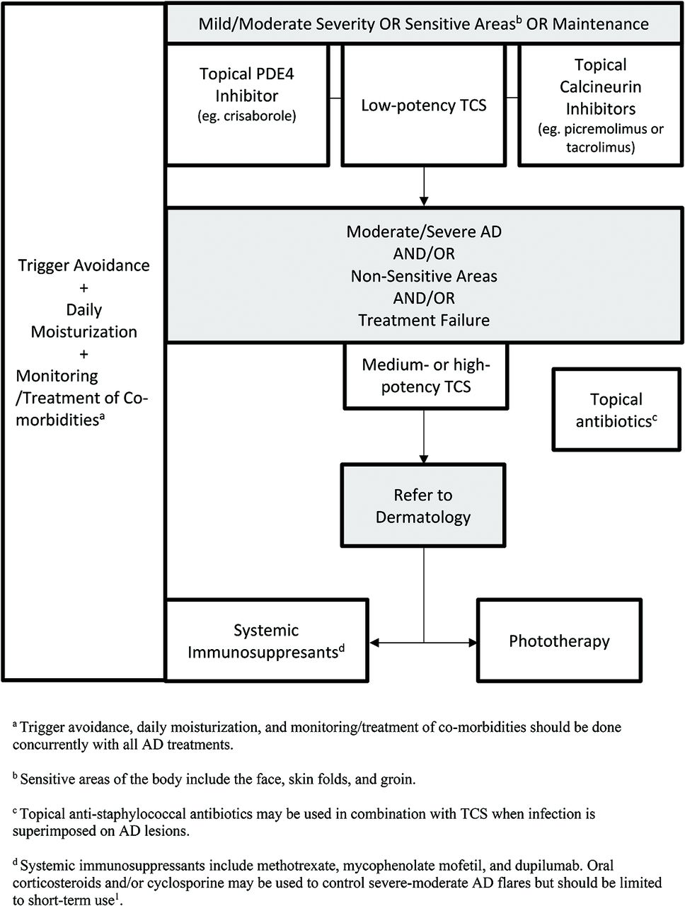
Treatment of AD. Abbreviations: TCS, Topical corticosteroids; AD, atopic dermatitis; PDE-4, Phosphodiesterase-4 inhibitor.

Trigger Avoidance

Some literature suggests that AD flares are seasonal and tend to correspond with periods of low humidity and low temperature.53 Patients with AD have a higher risk of irritant and allergic contact dermatitis, so common allergens such as fragranced products should be avoided.54,55 Clinicians should counsel patients on the importance of applying daily moisturizers to the entire body at least once per day and avoidance of common allergens and irritants.43

Topical Corticosteroids and Antibiotics

Topical corticosteroids (TCS) are the first-line medication56 for controlling mild-to-moderate AD (Table 3). Proper use involves considering both the potency of TCS and lesion location. Mild potency corticosteroids should be used on the face and skin folds and moderate potency corticosteroids on the body. They are typically applied twice daily for 2 to 4 weeks.39 Acute flares and thick/lichenified lesions on the body can be treated with high potency TCS (I, II) for up to 2 weeks but should then be tapered to a lower potency until the lesion resolves.39

View this table:
  • View inline
  • View popup
Table 3.

Prescription Therapies for Atopic Dermatitis

Adverse effects of TCS include cutaneous atrophy, especially on the face and skin folds.57 For these areas, low potency formulations (VI, VII) should be used56; however, if necessary, a 5- to 7-day course of high potency steroid tapered to a lower dose is acceptable.16 As a guideline, roughly 0.5 g (1 adult fingertip) of topical steroid should be spread over 2 palms worth of skin16 and used 1 to 2 times daily,58 with liberal concurrent emollient application.8,16 If no effects are seen after 2 weeks, clinicians should consider alternative potencies or medications or consider another diagnosis.16 Due to widespread concern regarding steroid atrophy among patients and health providers, many patients underapply TCS, which can lead to decreased efficacy and inadequate symptom control.55 Patients should be counseled on adequate application for the amount and duration of medication use. Prophylactic use of low-to-moderate potency TCS can be used to control continually refractory lesions.38 Further side effects of TCS, including telangiectasia, striae, acneiform or rosacea-like eruptions, allergic contact dermatitis, and impairment of wound healing, are uncommon with appropriate use and can be effectively mananged.59

Topical antibiotics, such as fusidic acid and mupirocin, are recommended as an adjunct medication with topical anti-inflammatories when signs of infection, such as crusting, oozing, and pus, coexist with AD.60 Ozenoxacin cream (1%) is a novel topical antibiotic with activity against MRSA that was recently approved for treating impetigo in patients ages 2 months and over and is applied twice daily for 5 days.61 Corticosteroid and antibiotic combination creams, such as 0.1% fusidic acid/betamethasone valerate or 1% fusidic acid/hydrocortisone exist, but the prophylactic use of topical antibiotics on AD lesions without signs of infection is not recommended.38 Swabs for culture and sensitivity should also be considered due to the rising incidence of resistant organisms.38 At present, antistaphylococcal topical antibiotics show noninferiority to 1 another for the treatment response of infected AD lesions.61

Topical Calcineurin Inhibitors

Topical calcineurin inhibitors (TCIs) are nonsteroidal agents that inhibit calcineurin-dependent T-cell activation.16 TCIs may be used as an alternative treatment or for prolonged treatment when steroid atrophy is a concern.38 They may also be used as adjunctive or maintenance therapy alongside high-dose TCS.16 Two TCIs are used in practice: 1% pimecrolimus and 0.03% or 0.1% tacrolimus, with both showing efficacy in short-term (3 to 12 week) and long-term (up to 12 months) use in adult and pediatric populations. Pimecrolimus (1%) is less effective than TCS but has value as a long-term maintenance therapy, and 0.1% tacrolimus is noninferior to moderately potent TCS.62 TCIs also may be used for the prophylactic treatment of recurrent flares, applied 2 to 3 times per week to prevent relapse.16 The most common side effect of TCIs16,62 is a burning or stinging sensation over the applied area; however, this often decreases with continual use.16 They should be discontinued if allergic/contact dermatitis or rosacea-like lesions develop.16 The US FDA has a black box warning concerning the long-term safety of TCIs due to an association of systemic immunosuppression with skin malignancies and lymphoma63; however, follow-up studies have failed to verify this.64,65 Currently, the Canadian Dermatology Association does not support this black box warning66 and has recently been removed from the product monograph of pimecrolimus.67

Topical PDE-4 Inhibitors

Crisaborole (2%) ointment is a topical PDE-4 inhibitor that was approved for AD treatment by Health Canada in 2018 for patients older than 2 years.67 It has shown benefit in controlling the signs and symptoms of AD in acute and chronic lesions for both adult and pediatric populations and is well-tolerated in areas susceptible to steroid atrophy, such as the face, skin folds, and groin.68 Current safety profiles of up to 52 weeks69 show that crisaborole has a reduced risk of systemic side effects compared with TCS and TCIs.70 The most common side effect noted in clinical trials has been temporary application site burning (4.4% crisaborole vs 1.2% control; P = .001) but typically resolves after the first day of application.68,70

Phototherapy

Phototherapy is a treatment option in AD for those in which topical treatments do not achieve adequate control.71 It typically involves a 2 to 5 times weekly administration of narrowband (309 to 312 nm) UVB72 to affected areas and shows benefit in the signs, symptoms, and quality of life in AD patients.73⇓⇓⇓⇓–78 Side effects of phototherapy range from erythema, advanced skin-aging, burning, itching, and nausea.16,78 Although it is effective, phototherapy access remains an issue for many patients and the frequency of treatments can be burdensome.

Dupilumab and JAK Inhibitors

Dupilumab is a subcutaneously administered human monoclonal antibody for the interleukin-4 receptor-α subunit and is approved for the treatment of moderate-severe AD in North America and Europe.79 A meta-analysis of 8 AD trials with dupilumab showed a relative risk of skin infection of 0.54 (95% CI, 0.42-0.70) and OD of eczema herpeticum of 0.34 (95% CI, 0.14-0.84) in dupilumab versus placebo, with no significant associations with herpes simplex virus or overall skin infections.80 Patients receiving dupilumab may also be at an increased risk of conjunctivitis.81 No baseline or follow-up laboratory monitoring is required. Ongoing studies are assessing longer-term effects.82

A new class of emerging AD therapy are JAK inhibitors, which act on a common inflammatory pathway of many dermatoses, including AD, alopecia areata, psoriasis, and vitiligo.83 Currently, phase II and III studies are ongoing for topical and oral formulations.

Conclusions

The management of AD should involve the liberal use of emollients and an avoidance of common irritants. In mild-to-moderate AD, TCS are the first-line medical therapy, followed by calcineurin inhibitors, which have been shown to be noninferior to TCS in controlling AD, without the risk of steroid atrophy. Topical PDE-4 inhibitors, such as crisaborole, are a new nonsteroidal anti-inflammatory approved for ages 2 years and older. For cases of AD that fail to be controlled with topical therapy, phototherapy should be considered, if available. Newer biologics, such as dupilumab, have been approved for moderate-to-severe AD and seem to have favorable efficacy and side-effect profiles according to randomized trials.

Notes

  • ↵# Co-first authors.

  • This article was externally peer reviewed.

  • Conflicts of Interest: PF has received honoraria and/or consulting fees and/or advisory board fees for AbbVie, Aralez, Altius, Cipher, Eli Lilly, Galderma, Janssen, Leo, Novartis, and Pfizer & Sanofi. YBY has no conflicts of interest to declare. CL has acted as a principal investigator, speaker and/or consultant and/or advisory board fees for AbbVie, Amgen AnatpysBio, Avillon, Arcutis, Bristol-Myers Squibb, Celgene Cipher Genentech, GlenMark, Incyte, Janssen, Leo Pharma, Kyowa, Pfizer, Merck Novartis, and Sanofi. BO has no conflicts of interest to declare. KOL has received honoraria and/or consulting fees and/or advisory board fees from Bausch, Eli Lilly & Pfizer.

  • Funding: None.

  • Biographical: PF is an Assistant Professor in the Division of Dermatology, University of Toronto. YBY is a senior medical student at the University of British Columbia. CL is an Associate Professor in the Division of Dermatology, University of Toronto. BO is an Assistant Professor in the Department of Family & Community Medicine, University of Toronto, North York General Hospital. KOL is a Lecturer in the Department of Family & Community Medicine, University of Toronto, St Michael's Hospital.

  • To see this article online, please go to: http://jabfm.org/content/33/4/626.full.

  • Received for publication December 12, 2019.
  • Revision received March 2, 2020.
  • Accepted for publication March 4, 2020.

References

  1. 1.↵
    1. Drucker A,
    2. Eyerich K,
    3. de Bruin-Weller M,
    4. et al
    . Use of systemic corticosteroids for atopic dermatitis: International Eczema Council consensus statement. Br J Dermatol 2018;178:768–75.
    OpenUrl
  2. 2.↵
    1. Barbarot S,
    2. Auziere S,
    3. Gadkari A,
    4. et al
    . Epidemiology of atopic dermatitis in adults: Results from an international survey. Allergy 2018;73:1284–1293.
    OpenUrl
  3. 3.↵
    1. Mallol J,
    2. Crane J,
    3. von Mutius E,
    4. et al
    . The International Study of Asthma and Allergies in Childhood (ISAAC) phase three: a global synthesis. Allergol Immunopathol (Madr) 2013;41:73–85.
    OpenUrlCrossRefPubMed
  4. 4.↵
    1. Williams H,
    2. Strachan D
    . The natural history of childhood eczema: observations from the British 1958 birth cohort study. Br J Dermatol 1998;139:834–9.
    OpenUrlPubMed
  5. 5.↵
    1. Silverberg JI,
    2. Hanifin JM
    . Adult eczema prevalence and associations with asthma and other health and demographic factors: a US population-based study. J Allergy Clin Immunol 2013;132:1132–8.
    OpenUrlCrossRef
  6. 6.↵
    1. Paternoster L,
    2. Standl M,
    3. Waage J,
    4. et al
    . Multi-ancestry genome-wide association study of 21,000 cases and 95,000 controls identifies new risk loci for atopic dermatitis. Nat Genet 2015;47:1449–56.
    OpenUrlCrossRefPubMed
  7. 7.↵
    1. Rodríguez E,
    2. Baurecht H,
    3. Herberich E,
    4. et al
    . Meta-analysis of filaggrin polymorphisms in eczema and asthma: robust risk factors in atopic disease. J Allergy Clin Immunol 2009;123:1361–70.e7.
    OpenUrlCrossRefPubMed
  8. 8.↵
    1. Weidinger S,
    2. Novak N
    . Atopic dermatitis. Lancet 2016;387:1109–22.
    OpenUrlCrossRefPubMed
  9. 9.↵
    1. Tsakok T,
    2. Woolf R,
    3. Smith C,
    4. Weidinger S,
    5. Flohr C
    . Atopic dermatitis: the skin barrier and beyond. Br J Dermatol 2019;180:464–74.
    OpenUrl
  10. 10.↵
    1. Kim J,
    2. Kim BE,
    3. Leung DY
    . Pathophysiology of atopic dermatitis: clinical implications. Allergy asthma Proc 2019;40:84–92.
    OpenUrl
  11. 11.↵
    1. Draaisma E,
    2. Garcia-Marcos L,
    3. Mallol J,
    4. et al
    . A multinational study to compare prevalence of atopic dermatitis in the first year of life. Pediatr Allergy Immunol 2015;26:359–66.
    OpenUrl
  12. 12.↵
    1. Kantor R,
    2. Kim A,
    3. Thyssen JP,
    4. Silverberg JI
    . Association of atopic dermatitis with smoking: a systematic review and meta-analysis. J Am Acad Dermatol 2016;75:1119–25.e1.
    OpenUrl
  13. 13.↵
    1. Schram M,
    2. Tedja A,
    3. Spijker R,
    4. Bos J,
    5. Williams H,
    6. Spuls PI
    . Is there a rural/urban gradient in the prevalence of eczema? A systematic review. Br J Dermatol 2010;162:964–73.
    OpenUrlCrossRefPubMed
  14. 14.↵
    1. Lee J,
    2. Seto D,
    3. Bielory L
    . Meta-analysis of clinical trials of probiotics for prevention and treatment of pediatric atopic dermatitis. J Allergy Clin Immunol 2008;121:116–21.e11.
    OpenUrlCrossRefPubMed
  15. 15.↵
    1. Pelucchi C,
    2. Chatenoud L,
    3. Turati F,
    4. et al
    . Probiotics supplementation during pregnancy or infancy for the prevention of atopic dermatitis: a meta-analysis. Epidemiology 2012;23:402–14.
    OpenUrlCrossRefPubMed
  16. 16.↵
    1. Fortson E,
    2. Feldman S,
    3. Strowd L
    . Management of atopic dermatitis: methods and challenges: New York (NY): Springer; 2017.
  17. 17.↵
    1. Silverberg JI,
    2. Margolis DJ,
    3. Boguniewicz M,
    4. et al
    . Distribution of atopic dermatitis lesions in United States adults. J Eur Acad Dermatol Venereol 2019;33:1341–8.
    OpenUrl
  18. 18.↵
    1. Hanifin J,
    2. Rajka G
    . Diagnostic features of atopic dermatitis. Dermatol Venereol 1980;92:44–7.
    OpenUrl
  19. 19.↵
    1. Rothe M,
    2. Grant-Kels J
    . Diagnostic criteria for atopic dermatitis. Lancet 1996;348:769–70.
    OpenUrlCrossRefPubMed
  20. 20.↵
    1. Weston W,
    2. Howe W
    . Overview of dermatitis (Eczema). UpToDate. Available from: https://www.uptodate.com/contents/overview-of-dermatitis-eczema. Published 2018.
  21. 21.↵
    Severity scoring of atopic dermatitis: the SCORAD Index (consensus report of the European Task Force on Atopic Dermatitis). Dermatology 1993;186:23–31.
    OpenUrlCrossRefPubMed
  22. 22.↵
    National Institute for Health and Clinical Excellence. Atopic eczema in under 12s: diagnosis and management. London: NICE; 2007. Available from: www.nice.org.uk/CG057.
  23. 23.↵
    1. Barrett M,
    2. Luu M
    . Differential diagnosis of atopic dermatitis. Immunol Allergy Clin North Am 2017;37:11–34.
    OpenUrl
  24. 24.↵
    1. Gupta A,
    2. Bluhm R
    . Seborrheic dermatitis. J Eur Acad Dermatol Venerol 2004;18:13–26.
    OpenUrlCrossRefPubMed
  25. 25.↵
    1. Johnston G,
    2. Sladden M
    . Scabies: diagnosis and treatment. BMJ 2005;331:619–22.
    OpenUrlFREE Full Text
  26. 26.↵
    1. Schlievert PM,
    2. Strandberg KL,
    3. Lin Y-C,
    4. Peterson ML,
    5. Leung DY
    . Secreted virulence factor comparison between methicillin-resistant and methicillin-sensitive Staphylococcus aureus, and its relevance to atopic dermatitis. J Allergy Clin Immunol 2010;125:39–49.
    OpenUrlCrossRefPubMed
  27. 27.↵
    1. Sun D,
    2. Ong PY
    . Infectious complications in atopic dermatitis. Immunol Allergy Clin North Am 2017;37:75–93.
    OpenUrlPubMed
  28. 28.↵
    1. Silverberg JI
    . Comorbidities and the impact of atopic dermatitis. Ann Allergy Asthma Immunol 2019;123:144–51.
    OpenUrl
  29. 29.↵
    1. Ascott A,
    2. Mulick A,
    3. Yu AM,
    4. et al
    . Atopic eczema and major cardiovascular outcomes: a systematic review and meta-analysis of population-based studies. J Allergy Clin Immunol 2019;143:1821–9.
    OpenUrl
  30. 30.↵
    1. Jeon C,
    2. Yan D,
    3. Nakamura M,
    4. et al
    . Frequency and management of sleep disturbance in adults with atopic dermatitis: a systematic review. Dermatol Ther (Heidelb) 2017;7:349–64.
    OpenUrl
  31. 31.↵
    1. Lee CH,
    2. Chuang HY,
    3. Hong CH,
    4. et al
    . Lifetime exposure to cigarette smoking and the development of adult-onset atopic dermatitis. Br J Dermatol 2011;164:483–489.
    OpenUrlPubMed
  32. 32.↵
    1. Silverberg JI,
    2. Song J,
    3. Pinto D,
    4. et al
    . Atopic dermatitis is associated with less physical activity in US adults. J Invest Dermatol 2016;136:1714–6.
    OpenUrl
  33. 33.↵
    1. Yaghmaie P,
    2. Koudelka CW,
    3. Simpson EL
    . Mental health comorbidity in patients with atopic dermatitis. J Allergy Clin Immunol 2013;131:428–33.
    OpenUrlCrossRefPubMed
  34. 34.↵
    1. Patel KR,
    2. Immaneni S,
    3. Singam V,
    4. Rastogi S,
    5. Silverberg JI
    . Association between atopic dermatitis, depression, and suicidal ideation: a systematic review and meta-analysis. J Am Acad Dermatol 2019;80:402–10.
    OpenUrl
  35. 35.↵
    1. Sandhu JK,
    2. Wu KK,
    3. Bui T-L,
    4. Armstrong AW
    . Association between atopic dermatitis and suicidality: a systematic review and meta-analysis. JAMA Dermatol 2019;155:178–87.
    OpenUrl
  36. 36.↵
    1. Schmitt J,
    2. Romanos M,
    3. Schmitt NM,
    4. Meurer M,
    5. Kirch W
    . Atopic eczema and attention-deficit/hyperactivity disorder in a population-based sample of children and adolescents. JAMA 2009;301:724–6.
    OpenUrlCrossRefPubMed
  37. 37.↵
    1. Chamlin SL,
    2. Mattson CL,
    3. Frieden IJ,
    4. et al
    . The price of pruritus: sleep disturbance and cosleeping in atopic dermatitis. Arch Pediatr Adolesc Med 2005;159:745–50.
    OpenUrlCrossRefPubMed
  38. 38.↵
    1. Weinstein M,
    2. Barber K,
    3. Bergman J,
    4. et al
    . Atopic dermatitis: a practical guide to management. Health Care Provid Resour 2016;12:1–12.
    OpenUrl
  39. 39.↵
    1. Dhadwal G,
    2. Albrecht L,
    3. Gniadecki R,
    4. et al
    . Approach to the assessment and management of adult patients with atopic dermatitis: a consensus document. section IV: treatment options for the management of atopic dermatitis. J Cutan Med and Surg 2018;22:21S–29S.
    OpenUrl
  40. 40.↵
    1. Draelos ZD
    . The science behind skin care: moisturizers. J Cosmet Dermatol 2018;17:138–44.
    OpenUrl
  41. 41.↵
    1. Feingold KR
    . Thematic review series: skin lipids. The role of epidermal lipids in cutaneous permeability barrier homeostasis. J Lipid Res 2007;48:2531–46.
    OpenUrlAbstract/FREE Full Text
  42. 42.↵
    1. Lowe A,
    2. Su J,
    3. Allen K,
    4. et al
    . A randomized trial of a barrier lipid replacement strategy for the prevention of atopic dermatitis and allergic sensitization: the PEBBLES pilot study. Br J Dermatol 2018;178:e19–e21.
    OpenUrl
  43. 43.↵
    1. Horimukai K,
    2. Morita K,
    3. Narita M,
    4. et al
    . Application of moisturizer to neonates prevents development of atopic dermatitis. J Allergy Clin Immunol 2014;134:824–30.e6.
    OpenUrlCrossRefPubMed
  44. 44.↵
    1. Simpson EL,
    2. Chalmers JR,
    3. Hanifin JM,
    4. et al
    . Emollient enhancement of the skin barrier from birth offers effective atopic dermatitis prevention. J Allergy Clin Immunol 2014;134:818–23.
    OpenUrlCrossRefPubMed
  45. 45.↵
    1. Evangelista MT,
    2. Abad-Casintahan F,
    3. Lopez-Villafuerte L
    . The effect of topical virgin coconut oil on SCORAD index, transepidermal water loss, and skin capacitance in mild to moderate pediatric atopic dermatitis: a randomized, double-blind, clinical trial. Int J Dermatol 2014;53:100–8.
    OpenUrl
  46. 46.↵
    1. Moro G,
    2. Arslanoglu S,
    3. Stahl B,
    4. et al
    . A mixture of prebiotic oligosaccharides reduces the incidence of atopic dermatitis during the first six months of age. Arch Dis Child 2006;91:814–9.
    OpenUrlAbstract/FREE Full Text
  47. 47.↵
    1. Grüber C,
    2. Van Stuijvenberg M,
    3. Mosca F,
    4. et al
    . Reduced occurrence of early atopic dermatitis because of immunoactive prebiotics among low-atopy-risk infants. J Allergy Clin Immunol 2010;126:791–7.
    OpenUrlCrossRefPubMed
  48. 48.↵
    1. Hon KL,
    2. Kung JSC,
    3. Ng WGG,
    4. et al
    . Emollient treatment of atopic dermatitis: latest evidence and clinical considerations. Drugs Context 2018;7:212530.
    OpenUrl
  49. 49.↵
    1. Hon KL,
    2. Leung AK,
    3. Barankin B
    . Barrier repair therapy in atopic dermatitis: an overview. Am J Clin Dermatol 2013;14:389–99.
    OpenUrlCrossRefPubMed
  50. 50.↵
    1. Wong S-M,
    2. Ng TG,
    3. Baba R
    . Efficacy and safety of sodium hypochlorite (bleach) baths in patients with moderate to severe atopic dermatitis in Malaysia. J Dermatol 2013;40:874–80.
    OpenUrlPubMed
  51. 51.↵
    1. Huang JT,
    2. Rademaker A,
    3. Paller AS
    . Dilute bleach baths for Staphylococcus aureus colonization in atopic dermatitis to decrease disease severity. Arch Dermatol 2011;147:246–7.
    OpenUrlCrossRefPubMed
  52. 52.↵
    1. Chopra R,
    2. Vakharia PP,
    3. Sacotte R,
    4. Silverberg JI
    . Efficacy of bleach baths in reducing severity of atopic dermatitis: a systematic review and meta-analysis. Ann Allergy Asthma Immunol 2017;119:435–40.
    OpenUrlPubMed
  53. 53.↵
    1. Engebretsen K,
    2. Johansen J,
    3. Kezic S,
    4. Linneberg A,
    5. Thyssen J
    . The effect of environmental humidity and temperature on skin barrier function and dermatitis. J Eur Acad Dermatol Venereol 2016;30:223–49.
    OpenUrl
  54. 54.↵
    1. Scheinman PL
    . Allergic contact dermatitis to fragrance: a review. Am J Contact Dermatitis 1996;7:65–76.
    OpenUrlPubMed
  55. 55.↵
    1. Larsen W,
    2. Nakayama H,
    3. Lindberg M
    . Fragrance contact dermatitis: a worldwide multicenter investigation (part I). Am J Contact Dermat 1996;7:77–83.
    OpenUrlCrossRefPubMed
  56. 56.↵
    1. Hoare C,
    2. Li Wan Po A,
    3. Williams H
    . Systematic review of treatments for atopic eczema. Health Technol Assess 2000;4:1.
    OpenUrlPubMed
  57. 57.↵
    1. Mayba JN,
    2. Gooderham MJ
    . Review of atopic dermatitis and topical therapies. J Cutan Med Surg 2017;21:227–36.
    OpenUrl
  58. 58.↵
    1. Green C,
    2. Colquitt J,
    3. Kirby J,
    4. Davidson P,
    5. Payne E
    . Clinical and cost-effectiveness of once-daily versus more frequent use of same potency topical corticosteroids for atopic eczema: a systematic review and economic evaluation. Health Technol Assess 2004;8:1–120.
    OpenUrlPubMed
  59. 59.↵
    1. Bewley A
    , Dermatology Working Group. Expert consensus: time for a change in the way we advise our patients to use topical corticosteroids. Br J Dermatol 2008;158:917–20.
    OpenUrlCrossRefPubMed
  60. 60.↵
    1. Rosen T,
    2. Albareda N,
    3. Rosenberg N,
    4. et al
    . Efficacy and safety of ozenoxacin cream for treatment of adult and pediatric patients with impetigo: a randomized clinical trial. JAMA Dermatol 2018;154:806–13.
    OpenUrl
  61. 61.↵
    1. Cowling T,
    2. Jones S
    . Topical antibiotics for infected wounds: a review of the clinical effectiveness and guidelines. Ottawa (ON): Canadian Agency for Drugs and Technologies in Health; 2017.
  62. 62.↵
    1. El-Batawy MMY,
    2. Bosseila M-W,
    3. Mashaly HM,
    4. Hafez V
    . Topical calcineurin inhibitors in atopic dermatitis: a systematic review and meta-analysis. J Dermatol Sci 2009;54:76–87.
    OpenUrlCrossRefPubMed
  63. 63.↵
    1. Legendre L,
    2. Barnetche T,
    3. Mazereeuw-Hautier J,
    4. Meyer N,
    5. Murrell D,
    6. Paul C
    . Risk of lymphoma in patients with atopic dermatitis and the role of topical treatment: A systematic review and meta-analysis. J Am Acad Dermatol 2015;72:992–1002.
    OpenUrlPubMed
  64. 64.↵
    Bausch Health Canada. Elidel (pimecrolimus) product monograph. Available from: https://pdf.hres.ca/dpd_pm/00053301.PDF. Published November 3, 2011. Accessed March 2, 2020.
  65. 65.↵
    1. Cury Martins J,
    2. Martins C,
    3. Aoki V,
    4. Gois A,
    5. Ishii H,
    6. da Silva E
    . Topical tacrolimus for atopic dermatitis. Cochrane Database Syst Rev 2015;2015:CD009864.
    OpenUrl
  66. 66.↵
    Canadian Dermatology Association. Topical calcineurin inhibitors position statement. Available from: https://www.dermatology.ca/tciposition-statement/. Published 2018.
  67. 67.↵
    1. Zebda R,
    2. Paller AS
    . Phosphodiesterase 4 inhibitors. J Am Acad Dermatol 2018;78:S43–S52.
    OpenUrl
  68. 68.↵
    1. Hoy SM
    . Crisaborole ointment 2%: a review in mild to moderate atopic dermatitis. Am J Clin Dermatol 2017;18:837–43.
    OpenUrl
  69. 69.↵
    1. Eichenfield LF,
    2. Call RS,
    3. Forsha DW,
    4. et al
    . Long-term safety of crisaborole ointment 2% in children and adults with mild to moderate atopic dermatitis. J Am Acad Dermatol 2017;77:641–9.e5.
    OpenUrlPubMed
  70. 70.↵
    1. Paller AS,
    2. Tom WL,
    3. Lebwohl MG,
    4. et al
    . Efficacy and safety of crisaborole ointment, a novel, nonsteroidal phosphodiesterase 4 (PDE4) inhibitor for the topical treatment of atopic dermatitis (AD) in children and adults. J Am Acad Dermatol 2016;75:494–503.e496.
    OpenUrlCrossRefPubMed
  71. 71.↵
    1. Sidbury R,
    2. Davis DM,
    3. Cohen DE,
    4. et al
    . Guidelines of care for the management of atopic dermatitis: section 3. Management and treatment with phototherapy and systemic agents. J Am Acad Dermatol 2014;71:327–49.
    OpenUrlCrossRefPubMed
  72. 72.↵
    1. Jekler J,
    2. Larkö O
    . UVB phototherapy of atopic dermatitis. Br J Dermatol 1988;119:697–705.
    OpenUrlCrossRefPubMed
  73. 73.↵
    1. Jekler J,
    2. Larkö O
    . Combined UVA-UVB versus UVB phototherapy for atopic dermatitis: a paired-comparison study. J Am Acad Dermatol 1990;22:49–53.
    OpenUrlCrossRefPubMed
  74. 74.↵
    1. Meduri NB,
    2. Vandergriff T,
    3. Rasmussen H,
    4. Jacobe H
    . Phototherapy in the management of atopic dermatitis: a systematic review. Photoderm Photoimm Photomed 2007;23:106–12.
    OpenUrl
  75. 75.↵
    1. Tzaneva S,
    2. Seeber A,
    3. Schwaiger M,
    4. Hönigsmann H,
    5. Tanew A
    . High-dose versus medium-dose UVA1 phototherapy for patients with severe generalized atopic dermatitis. J Am Acad Dermatol 2001;45:503–7.
    OpenUrlPubMed
  76. 76.↵
    1. Abeck D,
    2. Schmidt T,
    3. Fesq H,
    4. et al
    . Long-term efficacy of medium-dose UVA1 phototherapy in atopic dermatitis. J Am Acad Dermatol 2000;42:254–7.
    OpenUrlCrossRefPubMed
  77. 77.↵
    1. Grundmann-Kollmann M,
    2. Behrens S,
    3. Podda M,
    4. Peter RU,
    5. Kaufmann R,
    6. Kerscher M
    . Phototherapy for atopic eczema with narrow-band UVB. J Am Acad Dermatol 1999;40:995–7.
    OpenUrlPubMed
  78. 78.↵
    1. Sidbury R,
    2. Tom WL,
    3. Bergman JN,
    4. et al
    . Guidelines of care for the management of atopic dermatitis: Section 4. Prevention of disease flares and use of adjunctive therapies and approaches. J Am Acad Dermatol 2014;71:1218–33.
    OpenUrlCrossRefPubMed
  79. 79.↵
    1. Gooderham MJ,
    2. Hong H-h,
    3. Eshtiaghi P,
    4. Papp KA
    . Dupilumab: a review of its use in the treatment of atopic dermatitis. J Am Acad Dermatol 2018;78:S28–S36.
    OpenUrl
  80. 80.↵
    1. Fleming P,
    2. Drucker AM
    . Risk of infection in patients with atopic dermatitis treated with dupilumab: a meta-analysis of randomized controlled trials. J Am Acad Dermatol 2018;78:62–9. e61.
    OpenUrlCrossRef
  81. 81.↵
    1. Agnihotri G,
    2. Shi K,
    3. Lio PA
    . A clinician's guide to the recognition and management of dupilumab-associated conjunctivitis. Drugs R D 2019;19:311–8.
    OpenUrl
  82. 82.↵
    1. Worm M,
    2. Simpson EL,
    3. Thaçi D,
    4. et al
    . Efficacy and safety of multiple dupilumab dose regimens after initial successful treatment in patients with atopic dermatitis: a randomized clinical trial. JAMA Dermatol 2020;156:131–143.
    OpenUrl
  83. 83.↵
    1. Damsky W,
    2. King BA
    . JAK inhibitors in dermatology: the promise of a new drug class. J Am Acad Dermatol 2017;76:736–44.
    OpenUrl
PreviousNext
Back to top

In this issue

The Journal of the American Board of Family     Medicine: 33 (4)
The Journal of the American Board of Family Medicine
Vol. 33, Issue 4
July-August 2020
  • Table of Contents
  • Table of Contents (PDF)
  • Cover (PDF)
  • Index by author
  • Front Matter (PDF)
Print
Download PDF
Article Alerts
Sign In to Email Alerts with your Email Address
Email Article

Thank you for your interest in spreading the word on American Board of Family Medicine.

NOTE: We only request your email address so that the person you are recommending the page to knows that you wanted them to see it, and that it is not junk mail. We do not capture any email address.

Enter multiple addresses on separate lines or separate them with commas.
Diagnosis and Management of Atopic Dermatitis for Primary Care Providers
(Your Name) has sent you a message from American Board of Family Medicine
(Your Name) thought you would like to see the American Board of Family Medicine web site.
CAPTCHA
This question is for testing whether or not you are a human visitor and to prevent automated spam submissions.
2 + 10 =
Solve this simple math problem and enter the result. E.g. for 1+3, enter 4.
Citation Tools
Diagnosis and Management of Atopic Dermatitis for Primary Care Providers
Patrick Fleming, Yue Bo Yang, Charles Lynde, Braden O'Neill, Kyle O. Lee
The Journal of the American Board of Family Medicine Jul 2020, 33 (4) 626-635; DOI: 10.3122/jabfm.2020.04.190449

Citation Manager Formats

  • BibTeX
  • Bookends
  • EasyBib
  • EndNote (tagged)
  • EndNote 8 (xml)
  • Medlars
  • Mendeley
  • Papers
  • RefWorks Tagged
  • Ref Manager
  • RIS
  • Zotero
Share
Diagnosis and Management of Atopic Dermatitis for Primary Care Providers
Patrick Fleming, Yue Bo Yang, Charles Lynde, Braden O'Neill, Kyle O. Lee
The Journal of the American Board of Family Medicine Jul 2020, 33 (4) 626-635; DOI: 10.3122/jabfm.2020.04.190449
Twitter logo Facebook logo Mendeley logo
  • Tweet Widget
  • Facebook Like
  • Google Plus One

Jump to section

  • Article
    • Abstract
    • Background
    • Conclusions
    • Notes
    • References
  • Figures & Data
  • References
  • Info & Metrics
  • PDF

Related Articles

  • No related articles found.
  • PubMed
  • Google Scholar

Cited By...

  • The Most Frequently Read Articles of 2020
  • Medications, Medicating, and Medicated--When, Where, and How--Opioids and Others
  • Google Scholar

More in this TOC Section

  • Interpretating Normal Values and Reference Ranges for Laboratory Tests
  • Non-Surgical Management of Urinary Incontinence
  • Screening and Diagnosis of Type 2 Diabetes in Sickle Cell Disease
Show more Clinical Review

Similar Articles

Keywords

  • Atopic Dermatitis
  • Chronic Disease
  • Dermatology
  • Eczema
  • Primary Health Care
  • Quality of Life
  • Skin Diseases

Navigate

  • Home
  • Current Issue
  • Past Issues

Authors & Reviewers

  • Info For Authors
  • Info For Reviewers
  • Submit A Manuscript/Review

Other Services

  • Get Email Alerts
  • Classifieds
  • Reprints and Permissions

Other Resources

  • Forms
  • Contact Us
  • ABFM News

© 2025 American Board of Family Medicine

Powered by HighWire