Swollen lower limb—2: Lymphoedema
BMJ 2000; 320 doi: https://doi.org/10.1136/bmj.320.7248.1527 (Published 03 June 2000) Cite this as: BMJ 2000;320:1527
- Peter S Mortimer
Lymph conducting pathways may become reduced in number, obliterated, obstructed, or dysfunctional (because of failure of contractility or valve incompetence). A lack of sensitive methods for investigation makes it difficult to distinguish between these mechanisms. A defect in the lymph conducting pathways leads to primary lymphoedema; in practice this means no identifiable outside cause can be found. Secondary lymphoedema is due to factors originating outside the lymphatic system.

Primary lymphoedema
Congenital lymphoedema presenting at or soon after birth is rare. A family history suggests Milroy's disease. Swelling invariably affects both lower limbs, but the upper limbs and face may also swell.
Limb swelling may be the presenting and major manifestation of congenital lymphatic malformations either in a pure form—for example, diffuse lymphangioma—or in combination with a congenital vascular syndrome—for example, Klippel-Trenaunay syndrome (varicose veins, excessive long bone growth, and vascular birthmark).

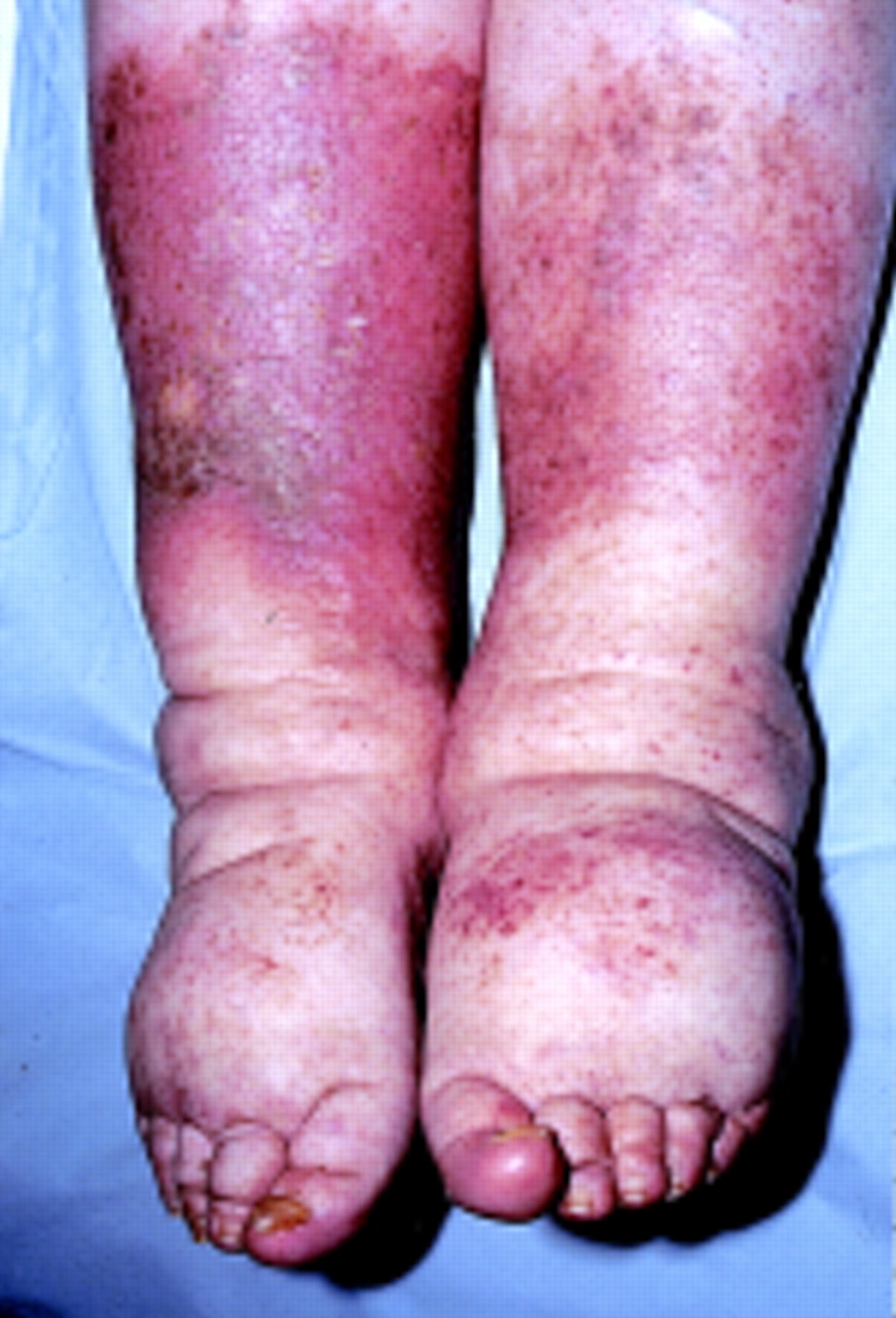
Primary lymphoedema with bilateral below knee swelling due to hypoplasia of peripheral lymphatic vessels
Most forms of primary lymphoedema present after puberty with foot and ankle swelling. Women are more often affected, and the condition may be familial—for example, Meige's disease. Lymph reflux due to lymphatic vessel hypertrophy or megalymphatics is clinically distinguishable.
Secondary lymphoedema
Lymphoedema manifesting with sudden onset of swelling of one whole leg suggests proximal obstruction. Pelvic causes of venous or lymphatic obstruction such as tumour or thrombosis must be excluded. In the Western world cancer treatment—for example, surgery or radiotherapy) is the commonest cause. Cancer itself rarely presents with lymphoedema except in advanced cases presenting late, such as prostate cancer, where venous obstruction may coexist. Relapsed tumour should always be considered in someone with limb swelling after apparent curative cancer treatment.

Kaposi-Stemmer sign: inability to pinch a fold of skin at base of second toe because of thickened skin indicates lymphoedema
Filariasis is probably the most common cause of secondary lymphoedema worldwide and should be considered in any patient with lymphoedema who has travelled or lived in an endemic area.
Clinical diagnosis of lymphoedema
The clinical diagnosis of lymphoedema depends on the history and characteristic skin changes. Although most swelling occurs in the subcutaneous layer, the skin becomes thicker (as demonstrated by the inablity to pinch a fold of skin at the base of the second toe), skin creases become enhanced, and a warty texture (hyperkeratosis) and papillomatosis develop. Such skin changes are termed “elephantiasis.”
The differential diagnosis includes venous oedema, “armchair legs,” and lipodystrophy or lipoedema, which is often misdiagnosed as lymphoedema.
Investigation of lymphoedema
Lymphoscintigraphy (isotope lymphography)
Lymphoscintigraphy is the best investigation for identifying oedema of lymphatic origin. Radiolabelled colloid or protein is injected into the first web space of each foot and monitored using a gamma camera as it moves to the draining lymph nodes. Measurement of tracer uptake within the lymph nodes after a defined interval will distinguish lymphoedema from oedema of non-lymphatic origin. The appearance of tracer outside the main lymph routes, particularly in the skin (dermal backflow), indicates lymph reflux and suggests proximal obstruction. Poor transit of isotope from the injection site suggests hypoplasia of the peripheral lymphatic system.

Dilatation of upper dermal lymphatics with consequent fibrosis gives rise to papillomatosis
Armchair legs (elephantiasis nostras verrucosis) develop in patients who sit in a chair day and night with their legs dependent. Patients with with cardiac or respiratory disease, stroke, spinal damage, or arthritis are predisoposed to this condition

Lipoedema only affects women and causes swelling between hip and ankle with sparing of the foot. The condition is symmetrical. The skin and subcutaneous tissues are soft and often tender with easy bruising

Lymphoscintigraphy. Radiolabelled colloid or protein is injected into the first web space of each foot and followed with a gamma camera as it moves to the draining lymph nodes. Tracer can be seen within the main lymphatic channels and lymph nodes as well as within the infection site. Collateral drainage is seen within the left thigh
Direct contrast x ray lymphography (lymphangiography)
After the lymph vessels have been identified with a vital dye, a contrast medium such as Lipiodol is administered directly into a peripheral lymphatic vessel, usually in the dorsum of the foot. In a normal limb the lymphangiogram will show opacification of five to 15 main collecting vessels as they converge on the lowermost inguinal lymph nodes. In patients with lymphatic obstruction the contrast medium will often reflux into the dermal network, so called “dermal backflow.”
Computed tomography and magnetic resonance imaging
Both computed tomography and magnetic resonance imaging detect a characteristic “honeycomb” pattern in the subcutaneous compartment that is not seen with other causes of oedema. In post-thrombotic syndrome the muscle compartment deep to the fascia is enlarged, whereas in lymphoedema it is unchanged. Thickening of the skin is also characteristic of lymphoedema, although it is not diagnostic. Magnetic resonance imaging is more informative than computed tomography because it can detect water.

Computed tomogram showing sections through normal thigh (left) and thigh with lymphoedema (right). Note thickened skin and honeycomb pattern
Management of lymphoedema
Most patients with lymphoedema are just told to live with it, but this is neither necessary nor acceptable.
Physical treatment to reduce swelling
Treatment is aimed at controlling lymph formation and improving lymph drainage through existing lymphatic vessels and collateral routes by applyingnormal physiological processes which stimulate lymph flow.
Physical treatment for lymphoedema
Prevention of infection
Prevention of acute inflammatory episodes (cellulitis or lymphangitis) is crucial because they can cause severe constitutional upset and deterioration in swelling. Care of the skin, good hygiene, control of skin diseases such as tinea pedis, and careful antiseptic dressings after minor wounds are all important. Antibiotics must be given promptly when an acute inflammatory episode occurs. In recurrent cellulitis the only effective treatment is prophylactic antibiotics—for example, phenoxymethylpenicillin 500 mg daily, for an indefinite period.
Drug treatment for lymphoedema
Diuretics are of little benefit in lymphoedema because their main action is to limit capillary filtration. Improvement in patients who are taking diuretics suggests that the predominant cause of the oedema is not lymphatic. The benefit of benzopyrones, such as coumarin or flavonoids, remains unproved.
Surgery
Surgery is of value in a few patients in whom the size and weight of a limb inhibit its use and interfere with mobility after physical treatment. Surgery is aimed at either removing excessive tissue (reducing or debulking operations)or bypassing local lymphatic defects.
Further reading
Footnotes
-
Peter S Mortimer is consultant skin physician, St George's Hospital and Royal Marsden Hospital, London.
-
The ABC of arterial and venous disease is edited by Richard Donnelly, professor of vascular medicine, University of Nottingham and Southern Derbyshire Acute Hospitals NHS Trust richard.donnelly{at}nottingham.ac.uk and Nick J M London, professor of surgery, University of Leicester, Leicester sms16{at}leicester.ac.uk.It will be published as a book later this year.